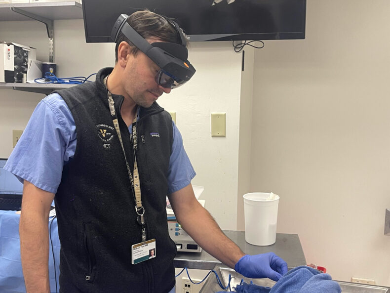
Last fall, the Vanderbilt Bill Wilkerson Center became the second facility in the country and third in the world to use a fully endoscopic surgical technique to remove an acoustic neuroma, a rare benign tumor on the balance and hearing nerves.
Alejandro Rivas, M.D., associate professor of Otolaryngology and Neurological Surgery, performed the surgery along with Reid Thompson, M.D., William F. Meacham Professor and chair of the Department of Neurological Surgery and professor of Otolaryngology. This is just one of the latest examples of Vanderbilt doctors employing an endoscopic technique that is less invasive than traditional surgical approaches.
“There are very few centers that are actually teaching their residents this technique,” Rivas said. “We are one of those centers in the United States and in the world.”
Traditionally, tumors of the ear are removed by drilling into the bone behind the ear and performing the surgery with a microscope. That is still the method used to remove large tumors, Rivas said. For smaller tumors, however, the endoscopic method offers an alternative.
An endoscope is a metal rod that has a lens at the end and fiber optics within, which projects a large, magnified image onto a screen. In tandem with surgical instruments, it allows for ear surgeries with minimal incision.
“From a patient’s perspective, there are a lot of advantages,” Rivas said. “It decreases pain. It’s a minimally invasive procedure. It has very similar, and in some cases better outcomes than using a traditional technique, with less post-operative care. It’s ideal for children, because, again, there are no incisions and children bounce back much faster.”
The endoscope also allows the surgeon to see through corners in cavities of the ear, improving visibility during surgery.
“Vanderbilt skull base surgeons are defining the future of surgery with these pioneering techniques,” Thompson said.
Ron Eavey, M.D., Guy M. Maness Professor and chair of Otolaryngology and director of the Vanderbilt Bill Wilkerson Center, said Rivas offers an annual continuing medical education (CME) course to teach other surgeons the endoscopic ear surgery method. “His course is one of a very few CME courses available on the subject, so it is international in reach and draws thought leaders to Vanderbilt.”
Besides removing tumors, other ear procedures are also benefiting from the endoscope. Removal of skin cysts (cholesteatomas) with endoscopes is very beneficial, as these lesions like to hide behind corners were the endoscope can reach, Rivas said. Perforations of the drum can also be corrected with endoscopic ear surgery.
Another example includes reconstruction of the tiny hearing bones when they are absent or do not move. Due to its enhanced vision, the endoscope allows perfect coupling between the drum and other ear structures when prostheses are used to reconstruct those missing bones.
Likewise, otosclerosis, which causes hearing loss as one of the ear bones (stapes) stops moving due to bone formation around it, can be addressed with endoscopic ear surgery. During surgery, the stapes can be replaced with prosthesis. The major advantage using the endoscope is in revision and with pediatric cases.
Elizabeth Pressey of Paris, Tennessee, who had been diagnosed with otosclerosis many years ago, came to see Rivas in December 2015 after failing multiple attempts with traditional ear surgery, trying to replace the stapes bone in her ear as well as repairing an eardrum, which often requires two separate surgeries. Rivas was able to do it in one surgery.
“He didn’t rush into anything at all,” she said. “He knew that he had a challenge ahead of him.”
She said Rivas’ technique was very successful.
“I hear now better than I ever have,” she said. “I had no pain whatsoever.”
Pressey had a follow-up visit with Rivas in November and everything looked good, she said, calling it a miracle.


