In the largest study of its kind, researchers at Vanderbilt University Medical Center have identified unexpected alterations in the exocrine tissues of the pancreas that occur in the two major forms of diabetes, and with aging and obesity.

Their work, published Oct. 26 in the journal Diabetes, represents a significant advance in understanding how Type 1 and Type 2 diabetes, generally considered diseases that affect primarily the insulin-producing endocrine cells of the pancreatic islet, also alter the digestive enzyme-secreting exocrine portion of the pancreas.
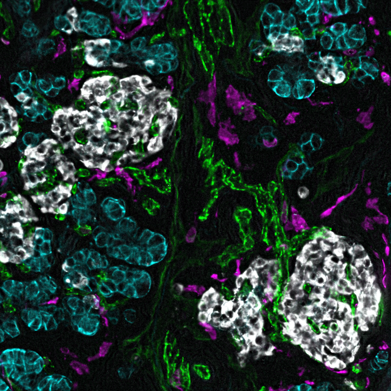
The great majority of the pancreas is exocrine tissue, with the pancreatic islets occupying only 1% – 2% of the organ.
Most diabetes research has focused on the endocrine pancreas, the islet cells, “because that’s where the physiological abnormalities arise,” said Jordan Wright, MD, PhD, instructor in Medicine and the paper’s co-first author with Adel Eskaros, MBBS, PhD. “There is increasing recognition that this is a disease of the whole pancreas.”
The pancreas has been very difficult to study because it is surrounded by other vital organs, hard to access, and it quickly deteriorates. “We almost never biopsy it,” Wright said. “Pancreas tissue for study must come from deceased donors or fragments of surgical specimens.”
Wright, an endocrinologist, and Eskaros, a clinical pathologist in the Division of Diabetes, Endocrinology, and Metabolism, and their colleagues led by senior authors Alvin C. Powers, MD, and Marcela Brissova, PhD, were able to study an unprecedently large and well-preserved group of tissue samples from 119 donors.
The tissue samples were provided by the Vanderbilt Pancreas Biorepository, the Human Pancreas Analysis Program of the National Institutes of Health (NIH), and the network for Pancreatic Organ Donors with Diabetes (nPOD). Twenty of the donors had Type 1 diabetes, 25 had Type 2 diabetes, and 74 had neither form of the disease.
Diabetes is characterized by high blood levels of glucose due to varying degrees of insulin deficiency, a hormone secreted by the pancreas that signals muscle, fat, and other organs to take up glucose and use it for energy.
Type 1 diabetes, which is often diagnosed in children, results from autoimmune destruction of the insulin-secreting endocrine cells, called beta cells, in the pancreas. Patients are taught to give themselves insulin, through shots or other means, to keep their glucose levels in check.
Type 2 diabetes, the most common form of the disease, often develops later in life and occurs when the pancreas fails to secrete enough insulin to compensate for the insulin insensitivity that is often associated with obesity. Treatment includes dietary changes and exercise, and oral or injectable diabetes medicines to help control blood glucose levels.
Control is important, because chronic high blood glucose levels increase the risk for heart and kidney disease, nerve and eye damage, and other serious complications. In addition to the disability and premature death caused by the disease, the cost of treating diabetes in the United States exceeds $325 billion a year.
That is why research to better understand what factors contribute to the disease is so important.
“As we set out to define the exocrine changes in diabetes, we quickly realized that tissues of the ‘normal’ human pancreas had not been well-described,” Wright said. “So, we first characterized how pancreatic tissues change across a wide range of donor ages and body weights.”
The researchers used this description of the “normal” pancreas to identify changes that are unique to both common forms of diabetes.
Pancreatic tissue from donors with Type 1 and Type 2 diabetes were significantly different in the extent of inflammation, atrophy, patterns of fibrosis and damage to blood vessels.
The researchers also noted changes to the exocrine tissues associated with aging and diabetes. “These changes occur in response to tissue injury and inflammation,” Eskaros said. “In the case of persistent injury caused by disease, they can carry a risk for progression to cancer.”
These observations, the researchers wrote, will guide future studies of exocrine-endocrine “crosstalk,” chronic pancreatic injury, and the risk of pancreatic cancer in diabetes. In particular, “we really hope to better understand what is driving those changes, and what is their significance for the disease,” Wright said.
“Exocrine tissues are hard to culture,” he said. “That’s a major challenge we’re trying to overcome … so we can study how they interact (with endocrine tissue). If you change the endocrine cells, what effect does that have on the exocrine cells, and vice versa.”
Brissova is research professor of Medicine. Powers is the Joe C. Davis Professor of Biomedical Science, professor of Medicine and of Molecular Physiology and Biophysics, director of the Vanderbilt Diabetes Center and of the Vanderbilt Diabetes Research and Training Center, and chief of the Division of Diabetes, Endocrinology, and Metabolism.
Other VUMC co-authors included Diane Saunders, PhD, research assistant professor of Medicine, Annika Windon, MD, assistant professor of Pathology, Microbiology and Immunology, Regina Jenkins, Amber Bradley, MS, Radhika Aramandla, MS, Sharon Philips, MSPH, and Hakmook Kang, PhD, associate professor of Biostatistics.
The research was supported by the Human Islet Research Network, Human Pancreas Analysis Program, NIH grants DK106755, DK123716, DK123743, DK120456, DK104211, DK108120, DK112232, DK112217, DK123594, DK133691, DK117147, EY032442, DK007061, and DK20593, the Leona M. and Harry B. Helmsley Charitable Trust, JDRF, Doris Duke Charitable Foundation, and U.S. Department of Veterans Affairs.
The content is solely the responsibility of the authors and does not necessarily represent the official views of the National Institutes of Health.