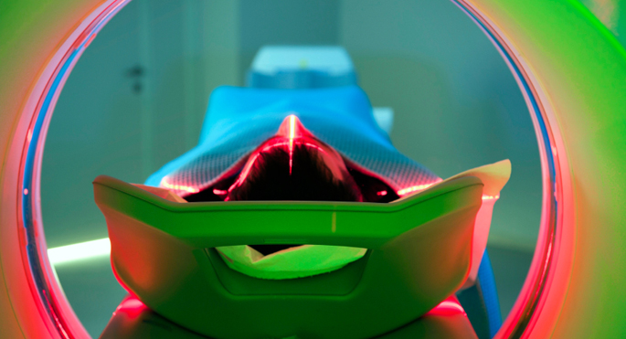
Researchers at Vanderbilt University Medical Center are investigating whether sonograms of fallopian tubes can be effective for the early detection of ovarian cancer, and their ongoing study shows promise.
Ovarian cancer is the most lethal form of gynecologic cancer because it is most often diagnosed at late stage. The symptoms are vague, and there is currently no screening protocol recommended for early detection. An interim study recently published in WFUMB Ultrasound Open, the official journal of the World Federation for Ultrasound in Medicine and Biology, provides an update on their progress.
The investigators shared data on four women who underwent ultrasounds with intraluminal contrast for improved imaging of the fallopian tube lumen, the interior portion of the organ. The four women carried the BRCA+ mutation and had scheduled risk reduction surgery for removal of their ovaries and fallopian tubes. Although biopsies after surgery revealed that none of the four women had cancerous lesions, one of the participants did have inflammation of the fallopian tube. The area of inflammation corresponded to the same spot where the ultrasound detected lower luminal circularity values relative to the adjacent segments of the tube and overall high rates of change of lumen diameter.

The finding supports the use of fallopian tube luminal contrast enhancement during ultrasounds to detect abnormalities.
“We are very excited about our findings thus far and look forward to further investigation on the role of fallopian tube imaging in early cancer detection,” said Jessica P. Miller, MD, PhD, a diagnostic radiologist and biomedical engineer, who is the lead researcher and corresponding author of the study.
The study is ongoing and continues to recruit participants as improvements are made to the imaging techniques and protocols for obtaining sonograms of the fallopian tube lumen. The researchers noted that one potential drawback is that their current technique may be limited in the evaluation of the fimbriae, the site where the contrast exits the fallopian tube and enters the peritoneum. The fimbriae location is also the site from which most serous tubal intraepithelial carcinoma lesions are thought to originate. However, additional testing of patients and the future identification of carcinomic lesions may indicate that contrast loss at the fimbriae does not inhibit adequate imaging.
The researchers are continuing to refine the imaging technology. They plan to add targeted visualization of the fallopian tube utilizing microbubble ultrasound contrast and are also designing algorithms for improved 3D image processing.
Other Vanderbilt authors on the study include Ryan G. Morrison, DO, Emily Mechling, Karen Tisdale, BS, RDMS, Katherine Frederick-Dyer, MD, Brannan Griffin, MD, Ben Park, MD, PhD, Lauren Prescott, MD, MPH, Marta Crispens, MD, Ronald Alvarez, MD, MBA, and Arthur Fleischer, MD.
The Vanderbilt researchers received support from the Susan Morrow Legacy Foundation and an AIUM Discovery Grant.