
microscopy Archive — Page 1 of 1


-
May 29, 2014
Pioneers of Discovery: Investigator taps into artistic side to reveal cells’ secrets
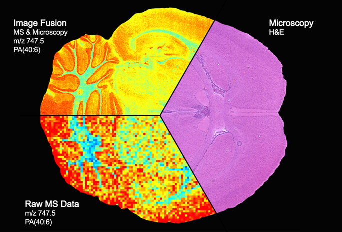
Dylan Burnette, Ph.D., points to one of the many striking photographs on his office walls. It’s a picture of a cell — a microscopic image showing yellow squiggles, bright purple lines and a turquoise oval on a black background, and it looks like abstract art.