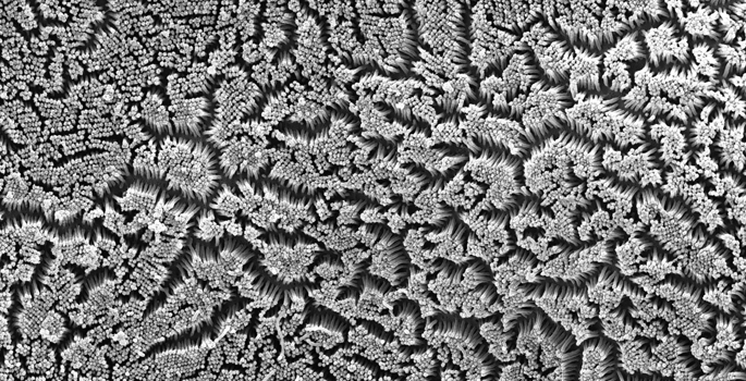
The epithelial cells that line organs like the intestines and kidneys build a special surface called the “brush border,” which consists of a dense array of finger-like microvilli. Matthew Tyska, Ph.D., and colleagues are exploring the molecular machinery that builds the border, which is critical for healthy organ function.
The investigators previously demonstrated that factors in an intermicrovillar adhesion complex (IMAC) stick the tips of microvilli together to build the border. However, it was unclear how the IMAC proteins became enriched at the microvillar tips.
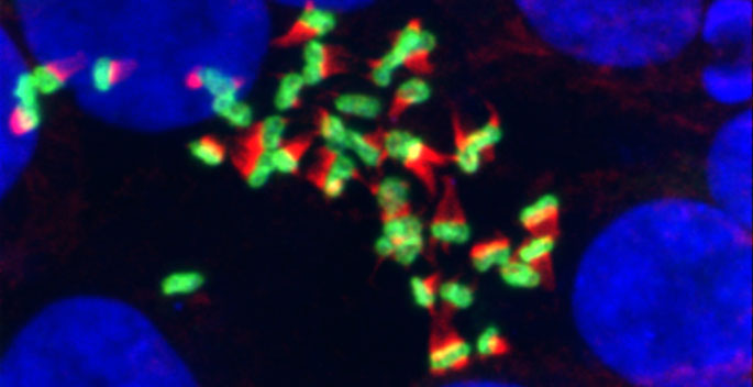
Now, in a report in Current Biology, the researchers demonstrate that the motor protein myosin-7b, which can move along the actin “core” of a microvillus, is required for normal microvillar organization. They showed that myosin-7b localizes to the tips of intestinal and kidney microvilli and that this localization requires a functional motor domain. Loss of myosin-7b impaired tip targeting of the IMAC factors.
The findings add to our understanding of brush border assembly in transporting and sensory epithelial tissues.
This research was supported by grants from the National Institutes of Health (grants DK075555, DK095811), by fellowship awards from the American Heart Association and the NIH (DK108528), and by the Vanderbilt Training Program in Stem Cell and Regenerative Developmental Biology.
Send suggestions for articles to highlight in Aliquots and any other feedback about the column to aliquots@vanderbilt.edu