
Matthew Tyska Archive — Page 1 of 1


-
September 13, 2018
How microvilli form
A protein called IRTKS helps build the microvilli that form the border of cells in the intestines, explaining why the protein is a frequent target of gut pathogens. -
January 31, 2018
Cell skeleton and the brush border
Vanderbilt researchers have discovered a role for microtubules — part of the cellular “skeleton” — in organizing the unique sidedness of the epithelial cells that line organs like the intestines. -
October 6, 2016
Motoring to the tips of the brush border
New findings implicate a motor protein in the assembly of the brush border in the intestines and kidneys – a specialized surface that is critical for healthy organ function. -
February 8, 2016
Building intestinal brush borders
Studies of the molecular complex that helps build specialized cellular surfaces could shed light on the mechanisms underlying a genetic deaf-blindness syndrome accompanied by intestinal disease. -
April 17, 2014
Nutrient-absorbing surface’s assembly revealed: study
Vanderbilt University researchers have discovered how intestinal cells build the "brush border" -- a specialized surface structure that is critical for absorbing nutrients and defending against pathogens. -
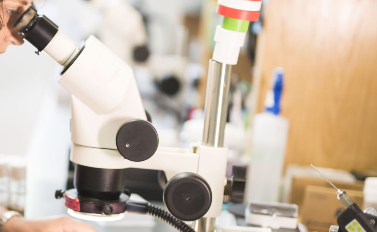
July 11, 2013
New ‘super’ microscopes sharpen cellular imaging
Two new “super-resolution” optical microscopes have put Vanderbilt University Medical Center on the cutting edge of cellular imaging, and are giving researchers their first views of the cell at the molecular level. -
May 14, 2012
Intestinal artillery launches anti-bacterial attack
The epithelial cells that line the intestines have a newly discovered mechanism for protecting us against microbes: they fire anti-bacterial "bullets" into the gut.