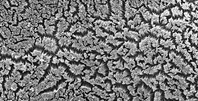
The epithelial cells lining organs like the intestines and kidneys build a special surface called the “brush border,” which consists of a dense array of finger-like protrusions.
Irina Kaverina, PhD, Matthew Tyska, PhD, and colleagues in Argentina explored the role of microtubules — part of the cellular “skeleton” — in building the border, which is critical for healthy organ function.
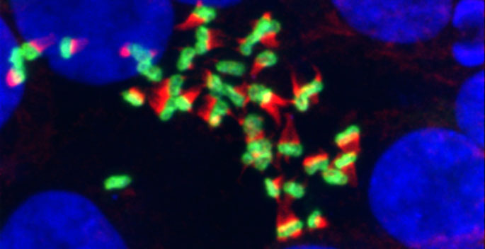
The investigators used a cell model of individual intestinal cell polarization (the establishment of “sidedness”) and found that disruption of microtubules prevented brush border formation. They confirmed the findings in cultured kidney cells.
They demonstrated that the microtubules essential for brush border development derived from the centrosome, not the Golgi, and that overexpression of a protein associated with microtubule plus ends facilitated brush border formation.
The findings, reported in the February issue of the Journal of Cellular Physiology, reveal a role for microtubules in the organization of the brush border and a novel mechanism of microtubule regulation of epithelial polarity.
This research was supported by CONICET, Agencia Nacional de Promoción Científica y Tecnológica, Argentina and by the National Institutes of Health (grants GM078373, DK106228, DK075555, DK095811).
Send suggestions for articles to highlight in Aliquots and any other feedback about the column to aliquots@vanderbilt.edu