A unique collaboration between imaging science, vascular medicine and rehabilitation at Vanderbilt University Medical Center is transforming the diagnosis and treatment of lipedema, a debilitating, abnormal deposition of fatty tissue that afflicts an estimated 17 million women in the United States.
Lipedema is often mistaken for obesity, yet it does not respond to diet or exercise. In women, the accumulation of fat occurs mainly in the legs, causing them “a lot of pain and difficulty with their daily activities,” said Aaron Aday, MD, MSc, assistant professor of Medicine.
“It’s a real thing,” said Aday, who specializes in vascular medicine. Yet many women “are having a horrible time, trying to find a diagnosis.”
About seven years ago, Rachelle Crescenzi, PhD, decided to do something about it. As a postdoctoral fellow in the Department of Radiology and Radiological Sciences at VUMC, she began to apply imaging techniques to improve diagnosis.
Lipedema “had been characterized in the 1940s, but people were relying on external measurements, which made it a very difficult diagnosis,” said Crescenzi, now an assistant professor in the department. With imaging, “we can look inside the body and show that this really is different from obesity.”
Lipedema is a disorder of the lymphatic system, which plays a major role in removing excess water (edema) from body tissues. Too much fluid in the heart, for example, can lead to heart failure.
Sodium (salt) plays a major role in regulating blood pressure and fluid volume. It is also a magnetic molecule and, as such, can be tracked by MRI.
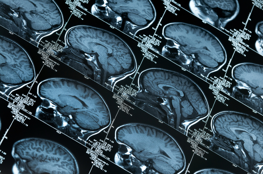
With funding from the Lipedema Foundation, Crescenzi and her colleagues developed an MRI strategy to quantify sodium content and fatty subcutaneous adipose tissue (SAT) throughout the body. They found that sodium and SAT volumes were significantly elevated in patients’ legs, but not arms, compared to women without lipedema.
In 2021 Crescenzi received the first R01 (independent investigator grant) ever awarded for lipedema research by the National Institutes of Health.
Supported by the five-year, $2.5 million grant, titled “Visualizing vascular mechanisms of lipedema,” Crescenzi established the Sodium Adipose and Lymphatics Translational (SALT) Lab, which is evaluating various treatments and diagnostic modalities.
In particular, the researchers are looking at conservative physical therapy to relieve patients’ lipedema-related leg pain, weakness, and excess fluid, improve mobility and optimize their home self-management program.
The manual techniques used in therapy include manual lymphatic drainage massage, myofascial and soft tissue releases to relax contracted muscles and surrounding connective tissues, along with use of graded negative pressure to expand and stretch tissue — all of which can improve lymphatic circulation.
This is where Paula Donahue, PT, DPT, MBA, comes in. Donahue, a Certified Lymphedema Therapist by the Lymphology Association of North America (CLT-LANA), is an assistant professor in the Department of Physical Medicine and Rehabilitation.
Working with Crescenzi’s team, they have shown that pain relief and improved function experienced by women with early lipedema after physical therapy correlated with a reduction of tissue sodium in the skin and SAT as measured by sodium and water MRI.
This proof-of-principle study, reported in August 2022 in the journal Lymphatic Research and Biology, showed that hands-on manual massage techniques not only helped patients feel better, but acted directly on the source of their pain.
One of the patients “was dealing with pain which was unexplained, and her doctor didn’t know what to do,” Donahue said.
With physical therapy, “her pain basically went down to zero and very quickly for her … For these individuals, it was very effective in changing their quality of life.”
“It wasn’t thought that you could compress out any of the pathologic tissue in lipedema, that it was just fat,” Crescenzi added. “But it really is fat and edema. We think sodium is a marker of inflammation … and it reduces after therapy.”
MRI is expensive, so Crescenzi and her colleagues are testing whether a lower-cost diagnostic method, ultrasound, is as effective in quantifying the extent of abnormal salt and SAT accumulation.
They have also developed a technique called magnetic resonance lymph angiography to better understand the hallmark features of lipedema. “We think the vascular system is just overloaded with a lot of edema, and it shows up on angiography,” Crescenzi said.
“We’re at the infancy of understanding this disease,” said Aday, co-author with Crescenzi and Donahue of the paper on the angiography technique published in June in the Journal of Magnetic Resonance Imaging. “We don’t have a good mechanistic understanding of this.”
The hope is that identifying what causes lipedema will lead to better ways to treat or prevent it.
“We’re in a position to develop a national resource for lipedema,” said Crescenzi, who presented the group’s latest findings in October at the American Vein and Lymphatic Society’s Annual Congress in New Orleans.
But it’s the patients who are driving the research and who are pushing for change in the treatment of lipedema. Said Crescenzi: “They taught me everything about it.”