
Imaging Archive — Page 1 of 13

-
May 20, 2026
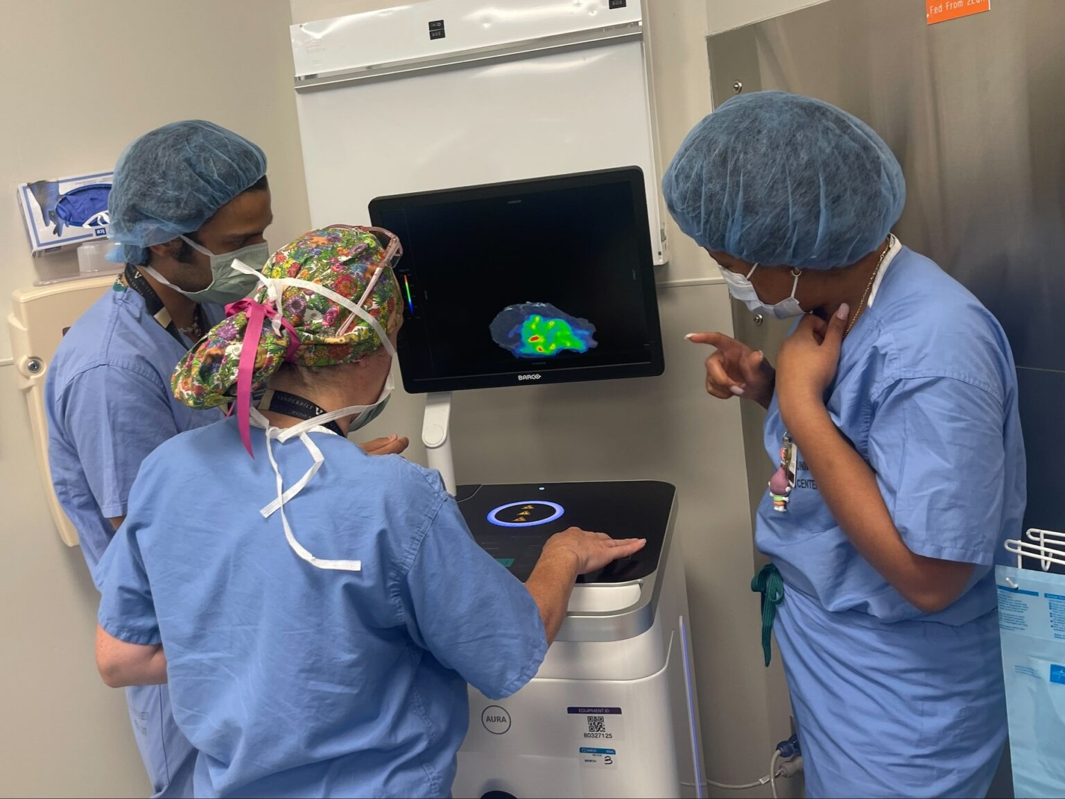
Neurosurgical ultrasound procedure simplifies care for patients with cerebrospinal fluid disorders
Ultrasound is easier on the brain, both physically and mentally. Reducing the burden for patients goes a long way toward a positive experience in the diagnosis and treatment process. -
April 9, 2026
How robotic bronchoscopy helped a Georgia man avoid unnecessary lung surgery
Oncologist Mohamed Shanshal, MD, said robotic bronchoscopy significantly improves getting patients the appropriate treatment as soon as possible, reducing their anxiety and improving care. -
April 7, 2026
Multi-institutional team pioneers retinal test for early Alzheimer’s disease
The potential for a retinal screening test was bolstered by a recent discovery that clearance of amyloid-beta oligomers by immune cells in retinal blood vessels was disrupted in donor eyes from individuals with Alzheimer’s disease, compared to age-matched, healthy control eyes. -
February 20, 2026
Ancient teeth, enduring patterns: Mummy study supports oral-cardiovascular disease link
CT scans of 37 Egyptian mummies found strong correlations between oral disease and calcified blood vessels, offering new evidence that the link between dental and cardiovascular health spans 4,000 years of human history. -
December 31, 2025
Vanderbilt’s Catherine Phillips is among ‘Forty Under 40’ radiologists for 2025
The online magazine called Philips “a visionary leader in academic radiology whose impact spans clinical excellence, operational innovation and educational advancement.” -
December 10, 2025
Study finds that meditation may help stimulate the brain’s waste removal system, providing restorative benefits like sleep
New research is informing how mindfulness meditation contributes to improvements in cognitive health and may have relevance to treating a range of degenerative brain conditions. -
November 18, 2025
Quantum computing team including Sunho Park wins competitive award
Their work focused on determining tissue and electrophysiological properties of the heart based on existing imaging approaches using quantum computing.