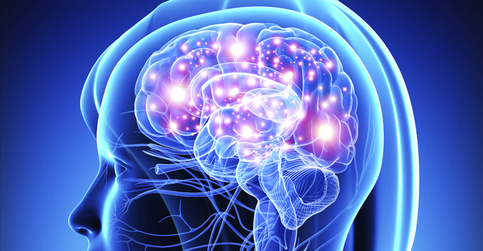
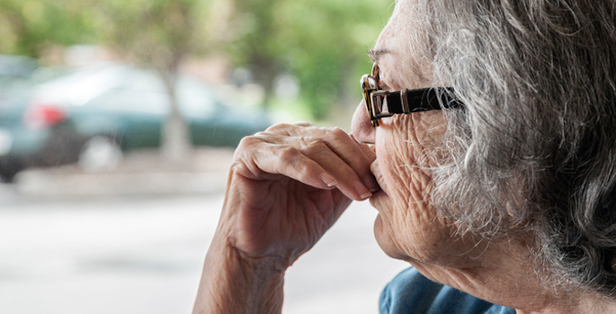
One’s quality of life tends to rest in the end on the ability to independently perform activities of daily living like shopping, preparing food and managing personal finances. In 324 participants aged 60 and older who were free of clinical dementia, MRI brain scans taken at study entry predicted independent function scores five years later. The study was reported by Corey Bolton, PsyD, Angela Jefferson, PhD, and colleagues in NeuroImage: Clinical.
The team gauged the predictive power of two neuroimaging results already known to be associated with functional status: the volume of gray matter in toto and in selected areas where the neurodegeneration seen in Alzheimer’s disease weighs most, and the amount of white matter hyperintensities (WMH), which are lesions indicating axonal loss and demyelination. Participants with less baseline gray matter and more baseline WHM experienced steeper declines in functional status.
The team also examined interactions of these biomarkers with cognitive status and with an Alzheimer’s-associated genetic variant.
The study data came from the Vanderbilt Memory and Aging Project at the Vanderbilt Memory and Alzheimer’s Center. Others on the study include Omair Khan, MS, Elizabeth Moore, MD, PhD, Kimberly Pechman, PhD, L. Taylor Davis, MD, Dandan Liu, PhD, Bennett Landman, PhD, Katherine Gifford, PsyD, and Timothy Hohman, PhD. The study was supported in part by the National Institutes of Health (AG034962).