


Imaging Archive — Page 12 of 14


-
October 1, 2014
Optical imaging in drug therapy screens
A non-invasive imaging tool could test potential cancer therapies quickly to personalize therapy for patients. -
September 24, 2014
Study finds accuracy of lung cancer imaging varies by region
A new analysis of published studies found that FDG-PET technology is less accurate in diagnosing lung cancer versus benign disease in regions where infections like histoplasmosis or tuberculosis are common. -
August 5, 2014
Technique brings spinal cord neural signaling into focus
Researchers in the Vanderbilt University Institute of Imaging Science have achieved the first conclusive non-invasive measurement of neural signaling in the spinal cords of healthy human volunteers. -
June 12, 2014
Powers helped launch ‘Nighthawk’ imaging service
Vanderbilt’s Thomas Powers, M.D., has learned to see exceptionally well in the dark. -
May 29, 2014
Pioneers of Discovery: Investigator taps into artistic side to reveal cells’ secrets
Dylan Burnette, Ph.D., points to one of the many striking photographs on his office walls. It’s a picture of a cell — a microscopic image showing yellow squiggles, bright purple lines and a turquoise oval on a black background, and it looks like abstract art. -
May 8, 2014
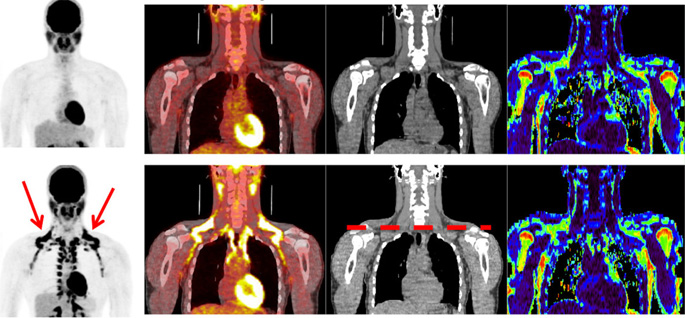
PET probe detects dying tumor cells
A novel PET imaging probe detects tumor cell death in vivo and could be useful for personalizing cancer medicine. -
February 28, 2014
Imaging guides Alzheimer gene search
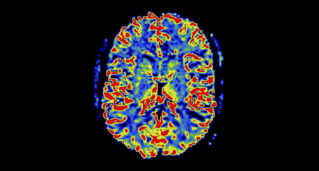
Using imaging data, Vanderbilt researchers discovered an association between a gene pair and brain changes in Alzheimer’s disease.