A team of surgeons and biomedical engineers at Vanderbilt University Medical Center and Vanderbilt University (VU) have shown the use of probe-based near infrared autofluorescence (NIRAF) technology helps confirm the identification of parathyroid glands during endocrine surgery, and their findings are part of a multisite trial of the technology now underway.

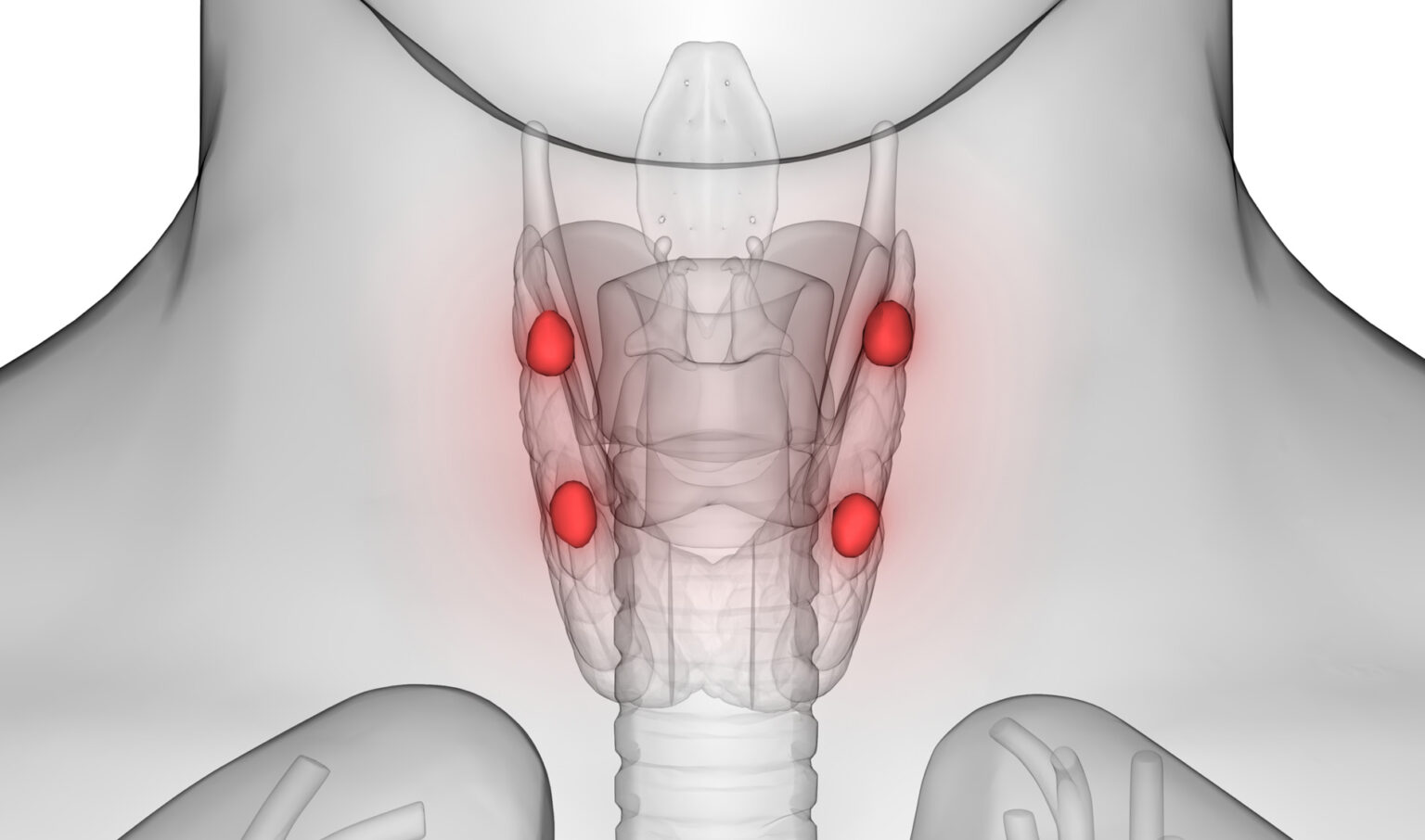
Parathyroid glands, which are about the size of a grain of rice, regulate blood calcium levels to support heart, nervous system, kidney and bone function. Locating parathyroid tissue and distinguishing it from other tissues during surgery is difficult, even for expert surgeons, because of the glands’ unpredictable anatomic position, small size and similar appearance to surrounding tissues. Preserving the delicate blood supply of parathyroid glands is also challenging.
In 2018, the Food and Drug Administration cleared for marketing two devices to assist with the detection of parathyroid glands in adults, including the probe-based device called the PTeye developed by a team at the Vanderbilt Biophotonics Center, led by Anita Mahadevan-Jansen, PhD, the Orrin H. Ingram Chair in Biomedical Engineering, and a VUMC endocrine surgery team led by Carmen Solórzano, MD, chair of the Department of Surgery, John L. Sawyers Professor of Surgical Sciences, and director of Vanderbilt Endocrine Surgery.
Parathyroids exhibit autofluorescence, meaning when the glands are illuminated with light of a specific wavelength, they reflect light back with a different wavelength. This allows the then glowing parathyroid glands to be more easily differentiated from other tissues in real time and without the need for frozen section biopsy to confirm parathyroid tissue, which can take 20-30 minutes.

In the recent randomized controlled trial, reported at the American Surgical Association and published in the journal Annals of Surgery, 160 patients undergoing parathyroidectomy (surgery to remove one or more parathyroid glands) were enrolled by a senior surgeon (more than 20 years’ experience) and a junior surgeon (less than five years’ experience) and randomly allocated to the probe-based NIRAF or control group.
Data collected included the number of parathyroids identified with high confidence, the number of frozen sections performed, surgery duration, and number of patients with persistent disease at the first post-operative visit.
“The clinical trial we performed at the Vanderbilt-Ingram Cancer Center and VUMC showed the device helped the surgeon feel more confident in their identification of parathyroids, more so than when the device was not used,” said Solórzano. “It also greatly reduced the need for frozen section biopsies, which is when you send a piece of tissue to the pathology lab to determine whether what you’re looking at is parathyroid tissue.”

In the probe group, the parathyroid identification rate of the senior surgeon improved from 3.2 to 3.6 parathyroids per patient, while that of the junior surgeon also rose from 2.2 to 2.5 parathyroids per patient. Parathyroid identification was even more prominent for surgeon trainees, increasing significantly from 0.9 to 2.9 parathyroids per patient. Additionally, there was a significant reduction in frozen sections in the probe group versus the control group.
Solórzano stressed that the technology is an adjunctive tool, intended to assist but not replace a surgeon’s visual identification of the parathyroid gland and frozen sections to confirm thyroid tissue type.
“I use this technology in my difficult surgical procedures, and I find that it makes a difference for my patients,” she said.
While thyroid surgery is generally a safe surgery with a low rate of complications, recent studies show that accidental damage or removal of healthy parathyroid glands occurs in 5-15% of patients. This leads to complications such as post-surgical hypoparathyroidism, or low levels of parathyroid hormone in the blood which causes hypocalcemia, or lower than normal levels of calcium.
Treatment for hypoparathyroidism is lifelong supplementation with calcium and vitamin D and the condition is associated with impaired quality of life and long-term renal complications. In patients with a diseased parathyroid gland, failure to find and remove the diseased tissue can lead to costly repeat surgeries and complications. Damage or removal of calcium-regulating parathyroid glands during endocrine surgery can put children at risk for poor growth and slow mental development.
“The most common surgeries in the head and neck that are endocrine related are thyroid and parathyroid procedures,” said Solórzano. “There are other procedures, too — for instance revision surgeries for patients who had thyroid cancer and now have come back with metastases in the lymph nodes or for patients who had a failed parathyroid surgery in the past.
“Our hope is that this technology can help improve identification and prevent the inadvertent resection of these parathyroid glands during surgery to remove cancer or benign tumors and thus prevent hypoparathyroidism. This is exciting for the pediatric population, too, because though these surgeries are not as common in this group, the parathyroids are tiny in children. So, really any help will move the needle toward fewer complications.”
A multicenter study comparing outcomes when using the probe-based NIRAF technology and not using the technology is underway for thyroid and parathyroid procedures with the University of Michigan, University of California-San Francisco, and the Medical College of Wisconsin joining VUMC.
In 2024, VUMC and Vanderbilt are also hosting the International Society of Innovative Technologies for Endocrine Surgery 6th Symposium ( https://isitesurgery.org/) in Nashville, during which advances to improve surgical outcomes, including the PTeye, will be showcased.
Additional collaborators for the study published in the Annals of Surgery include Colleen Kiernan, MD, MPH, Giju Thomas, PhD, Anuradha Patel, MD, Run Fan, PhD, MS, Fei Ye, PhD, MSPH, Parker Willmon, MS and Naira Baregamian, MD.