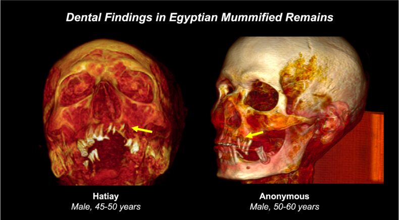
 Three-dimensional reconstructed CT scans of skulls from two named Egyptian mummies acquired by the Horus study. (illustration by Atlee Witt)

When it comes to our teeth and hearts, ancient mummies have a lot to tell us.
From an analysis of computed tomography (CT) scans of 37 Egyptian mummies, researchers at Vanderbilt Health and their colleagues determined that dental cavities and periodontal disease were highly prevalent in this ancient cohort and correlated with evidence of calcified blood vessels, an enduring marker of cardiovascular disease.

Their findings, reported Jan. 30 in the Journal of the American Dental Association, suggest that mummified teeth contain useful information for understanding the link between oral health and cardiovascular disease and, more broadly, that clues to improving the health of modern humans can be found in ancient bones.
“Atherosclerosis and dental disease both appear to be inflammatory processes linked with chronological age, in people who live today and in people who lived 4,000 years ago in Egypt,” said Katherine Van Schaik, MD, PhD, MA, assistant professor of Radiology & Radiological Sciences at Vanderbilt Health and the paper’s corresponding author.
“On the surface, this seems obvious — but why should this necessarily be the case, given modern advances in hygiene and medicine?” she asked. “Studies like this can help guide mechanistically focused assessments of the biology of aging.
“Historical remains provide a unique opportunity to study human biology apart from modern interventions. Evaluation of the biology of human aging and disease processes at time points 4,000 years apart can shed light on the most fundamental aspects of those mechanisms.”
Van Schaik is principal investigator of the Vanderbilt Program in Health over Time in the Vanderbilt Center for Antibody Therapeutics. The program studies longevity over the span of human history, from markers of disease preserved in mummified skeletal remains to the immune systems of modern patients.
The association between oral health and cardiovascular disease is well known.
Tooth decay and periodontal disease contribute to chronic inflammation and bacterial infections that in turn can contribute to atherosclerosis, a buildup of fat, cholesterol and calcium deposits in artery walls which can decrease blood flow to the heart, brain and vital organs.
While soft tissue deteriorates rapidly after death, teeth are a particularly well-preserved part of the skeleton, and modern imaging methods can detect the telltale tracks of vascular calcium deposits.
The current study, one of the first to correlate cardiovascular and dental disease in mummified remains, analyzed CT scans of 14 mummies identified as female and 23 identified as male. Their estimated ages at the time of death ranged from 19 to 60.
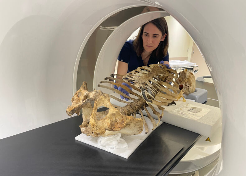
The CT images were acquired by the Horus team, an international, interdisciplinary research consortium named for the ancient Egyptian deity that represents kingship, healing, protection, the sun and the sky. Van Schaik is a current member of the group.
In addition to Van Schaik, the images for this study were analyzed by Atlee Witt, an MD/PhD student at Vanderbilt University School of Medicine and the paper’s first author, and by Derek Smith, DDS, PhD, a dentist and Vanderbilt-trained biostatistician who currently directs the Division of Biostatistics and Computational Biology at the University of Iowa College of Dentistry.
Among the 37 mummies, the men were less likely than the women to have calcified vascular beds. Today, just the opposite is observed — the severity of atherosclerosis and periodontitis is less pronounced in women.
While further research on the effect of sex on disease incidence in mummies is warranted, these findings support the value of studying “health over time,” Van Schaik said.