epithelial Archive — Page 1 of 1

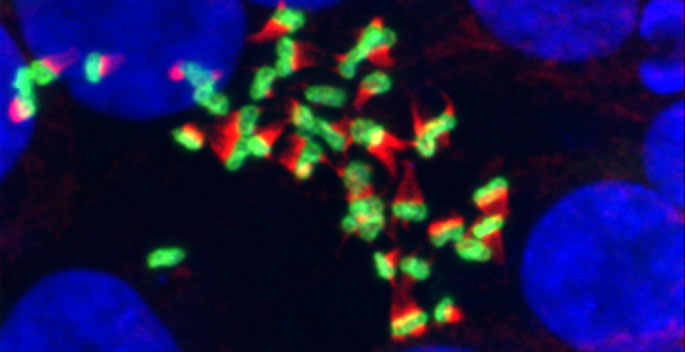
Fluorescent multicolored microscopic structures resembling clustered crystals or fibers against a black background.
September 19, 2019

Microvilli in motion

Live cell imaging studies have revealed that microvilli — finger-like protrusions on the surface of epithelial cells — move and collide as they form the brush border.

Fluorescence microscopy image showing blue-stained cell nuclei surrounded by green cell bodies and branching extensions against a black background; the cells appear separated and variably spread.
February 28, 2019

Protein loss promotes cell migration

The protein kinase STK17A plays a novel role in epithelial cells and its loss may contribute to colorectal cancer invasion and metastasis, Vanderbilt researchers report.

January 31, 2018

Cell skeleton and the brush border

Vanderbilt researchers have discovered a role for microtubules — part of the cellular “skeleton” — in organizing the unique sidedness of the epithelial cells that line organs like the intestines.

Gray-haired man in a striped shirt and tie sits in a laboratory beside a computer and illuminated research equipment.
November 29, 2012

Polarity protein suppresses tumor growth, invasion

The machinery that builds the characteristic shape of epithelial cells suppresses breast cancer formation and metastasis in a mouse model.

Fluorescence microscopy image showing blue circular structures and clustered green and red fluorescent signals against a dark background. Metadata text: “ImageJ=1.44e” and “unit=um.”
May 14, 2012

Intestinal artillery launches anti-bacterial attack

The epithelial cells that line the intestines have a newly discovered mechanism for protecting us against microbes: they fire anti-bacterial “bullets” into the gut.

March 18, 2011

Protein combos keep cells straight

Researchers define the protein interactions that establish our organs’ lining.