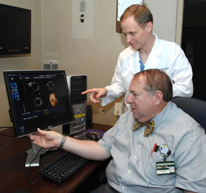
Arthur Fleischer, M.D., foreground, Andrej Lyshchik, M.D., Ph.D., and colleagues are studying a new imaging technique that may be able to spot ovarian cancer tumors at an earlier stage. (photo by Neil Brake)
Spotting ovarian cancer earlier goal of new imaging technique
It is not uncommon for growths to develop on female ovaries, but until recently physicians couldn't determine if the growth was a benign cyst or a cancerous tumor without an invasive procedure like surgery.
Now, a group of Vanderbilt-Ingram Cancer Center researchers, led by Arthur Fleischer, M.D., professor of Radiology and Radiological Sciences, has developed a promising new imaging tool which may eventually be used to identify ovarian cancer at an earlier stage. The study was published in the July issue of the Journal of Ultrasound in Medicine.
In normal tissue there is an orderly branching of blood vessels. But tumors develop a microscopic web of capillaries that are abnormal, with unique blood flow patterns and flow dynamics.
So Fleischer, working with Andrej Lyshchik, M.D., Ph.D., and Howard Jones, M.D., has been developing an intravenous contrast agent using inert microbubbles that are smaller than a red blood cell to highlight these changes.
“It starts with a solution that gets shaken up and produces the microbubbles,” Fleischer explained. “We inject the bubbles into an arm vein, followed by a small volume of normal saline. Then we use a specially tuned transvaginal sonographic probe to track the progress of those microbubbles as they light up the screen — it turns white when there is a network of abnormal blood vessels in a tumor. We can see the difference between vascularity in normal tissue and abnormal vascularity in tumors.”
In the study, supported by a grant from the National Institutes of Health and the National Cancer Institute, researchers tested this technique in 17 patients with 23 distinct ovarian masses and found statistically significant differences in microvessel flow between benign and malignant ovarian tumors. All of the women in the study underwent surgery and had their ovaries removed for further examination of the suspect tissue.
“One hundred percent of the malignant tumors and 43 percent of the benign lesions showed detectable contrast enhancement following the injection,” said Fleischer.
“This technique should become extensively utilized as a means to detect ovarian cancer in its earliest stage.”
The use of this new imaging tool for suspicious masses could save lives because ovarian cancer is often discovered at a late stage when it is notoriously difficult to treat. This silent disease, with subtle symptoms including abdominal swelling or intestinal distress, is not the most common gynecological cancer, but it is the most deadly, claiming approximately 15,280 lives in 2007, according to the National Cancer Institute.
Fleischer and his colleagues note that the study included a small patient sample and requires further investigation.
If additional studies validate this imaging technique, women whose masses appear to be benign may be spared from undergoing surgery, whereas those with malignancies will be caught at an earlier stage.


