More than a decade after a Vanderbilt University biomedical engineer discovered that the human parathyroid gland glows under near-infrared light, a Food and Drug Administration (FDA)-cleared device based on that finding will be used in a multisite clinical trial to further validate its efficacy.
“In the past, surgeons have had to rely on their visual identification of parathyroid glands,” said Anita Mahadevan-Jansen, PhD, Orrin H. Ingram Professor of Biomedical Engineering and director of the Vanderbilt Biophotonics Center. “Other techniques like ultrasound or sestamibi scans tend to work only when the parathyroid is diseased. It is gratifying to have our laboratory’s work culminate in clinically translatable science that can be used to improve patient care.”
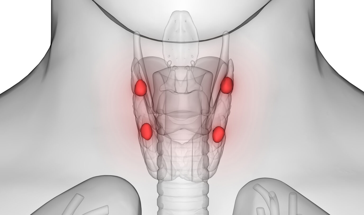
Parathyroid glands regulate blood calcium levels to support heart, nervous system, kidney and bone function. They are normally about the size of a grain of rice and found beside the larger thyroid gland. Locating parathyroid tissue and distinguishing it from nearby tissues during surgery can be challenging, even for expert surgeons.
In up to 20% of the 150,000 thyroid surgeries performed in the U.S. annually, healthy parathyroid glands are accidentally damaged or removed, leading to complications such as post-surgical hypocalcemia, or lower than normal levels of calcium. In patients with a diseased parathyroid gland, failure to find and remove the diseased tissue can lead to costly repeat surgeries and complications.
“About 20 million Americans have some form of thyroid disease, and more than 50,000 cases of thyroid cancer are diagnosed annually,” said Carmen Solórzano, MD, chair of the Department of Surgery and director of Vanderbilt Endocrine Surgery. “The PTeye system requires no contrast dye, can be used with normal operating room lighting, is non-invasive and has already proven to be very accurate. Our surgical group has worked closely with VU’s biomedical engineers on this technology, and we look forward to additional trials with the goal of improving surgical precision and improving outcomes for patients not just here, but throughout the world.”
The discovery of the parathyroid gland’s fluorescence was purely by chance. In 2008, Lisa White, MD, then a VUMC general surgery resident, noted the difficulty of locating the parathyroid gland during endocrine surgery. She contacted Mahadevan-Jansen who investigates the use of light-based technologies for biomedical applications to see if her research could be applied to the problem. Given that light often interacts uniquely with different tissues, these technologies have been used to aid in detecting disease and guiding tumor surgery.
As White worked alongside Mahadevan-Jansen and lab members, they discovered that parathyroid glands exhibit a higher degree of near infrared autofluorescence compared to adjacent neck tissues. Based on this finding, Mahadevan-Jansen and White theorized that detecting near infrared autofluorescence from parathyroid glands can be a way to reliably locate the glands during surgeries.
Mahadevan-Jansen and her team went further to develop PTeye, a hand-held, probe-based device which provides real-time guidance in identifying the parathyroid gland during surgery. When parathyroid tissue is detected, the system provides an audio signal and visual display. A device prototype was tested in a clinical study conducted in 2017 at VUMC and Ohio State University Medical Center. The results were published in the journals Thyroid, Surgery and the Journal of the American College of Surgeons.
During the study, the system accurately identified parathyroid glands 96.1% of the time, which led to FDA clearance of the device in November 2018. AiBiomed, a spinoff of Anasys Instruments Corp. (later acquired by Bruker Corporation), holds the license to produce and market the device. The PTeye recently received a 2019 R&D 100 Award from R&D World Magazine, as selected by a panel of expert judges.
The research team, led by principal investigator Mahadevan-Jansen, co-principal investigator Solórzano and Melinda Sanders, MD, received a National Institutes of Health (NIH) grant to support development of the device and evaluate its impact on surgery outcomes. Following VUMC Institutional Review Board approval, Solórzano is beginning a clinical trial to further assess the benefit of the PTeye device during surgeries, and several medical centers across the U.S. are expected to join a multisite study evaluating the clinical performance of PTeye.
A second FDA-cleared device, the Fluobeam 800 Clinic Imaging Device now marketed by Fluoptics, also resulted from Mahadevan-Jansen’s work. Fluobeam is an integrated fluorescence imaging device with a Class 1 laser as the light source and a near-infrared-sensitive camera. Compared to PTeye, Fluobeam localizes parathyroid glands by using its camera to capture images or videos of near infrared autofluorescence from the glands.
The Vanderbilt Center for Technology Transfer and Commercialization (CTTC) worked with Mahadevan-Jansen and Ai Biomed in the intellectual property and regulatory processes. This research was supported by the National Cancer Institute of the National Institutes of Health under award number R01CA212147.