A Vanderbilt University Medical Center interim analysis shows that there appears to be a considerable length of time before a subset of people develop inflammatory, scarring lung diseases where there is radiologically detectable evidence they will develop lung disease.
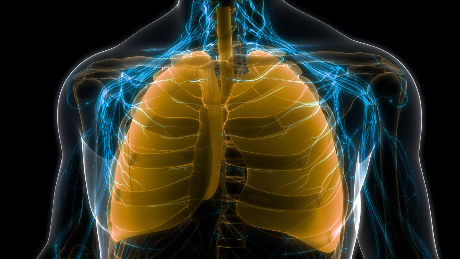
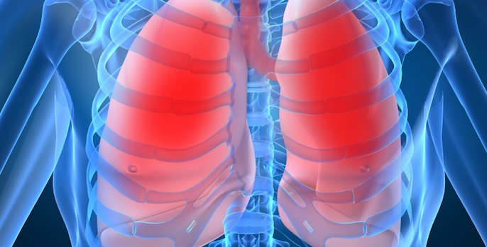
The interim analysis looked at a group of people who have at least two family members with Interstitial Lung Disease (ILD), a collection of numerous individual diseases that can cause both inflammation and scarring of lung tissue, particularly in those areas responsible for moving oxygen from the air into the bloodstream.
The family members enrolled in the study were not known to have lung disease nor symptoms. Most people in the study had relatives with pulmonary fibrosis (the scarring type of ILD).

“We were interested in looking at what proportion of those individuals without any symptoms or any known lung problems had early signs of disease on a screening CT scan of the lungs,” said Margaret Salisbury, MD, assistant professor of Medicine and the lead author of the study that appears online in the American Journal of Respiratory and Critical Care Medicine.
“We were also interested in identifying factors in people’s environments that might be risk factors for early disease on the screening CT scan,” she said.
The VUMC study looked at 336 family members who received CT scans and 265 people who completed exposure questionnaires.
The CT scans were done between late 2008 and early 2019. Subtle, suspicious findings on the scans were marked by a radiologist so that follow-up could determine whether they were getting worse over time and would go on to progress to pulmonary fibrosis.
A subgroup of the people enrolled early were able to get a follow-up CT scan five years later to determine whether there had been any progression of the early findings on the CT scan.
Twenty-three percent of the total number enrolled showed changes that could be early disease, Salisbury said. Of those having the second CT scan five years later, about 20% overall showed disease progression.
“In those with suspicious findings on their first CT scan, about 60% got worse,” Salisbury said. “In those with no suspicious findings, only about 6% developed changes,” she said.
The investigators also found that there were four environmental exposures on the questionnaire that were statistically significant with having early changes on the CT screening scan, Salisbury said.
“These four factors — mold contamination in the environment, exposure to birds (raising birds on a farm or keeping pet birds), exposure to lead and aluminum smelting in the workplace — are likely risk factors for early disease,” she said.
It’s already known that cigarette smoking is a very strong risk factor in developing these early changes as well as lung disease itself, she added.
Salisbury said the study is ongoing.
“We hope to get more information about the rate of disease development and progression as more people return and get their follow-up CT scans every five years. We’re also looking into ways we can learn how these environmental factors contribute to disease progression.
“The fact that a lot of people with early changes do progress suggests that even very minor findings on the CT scan might be meaningful and some of these environmental factors could be avoided. That’s something to keep in mind for people who have a family history of lung disease who might be at high risk,” Salisbury said.