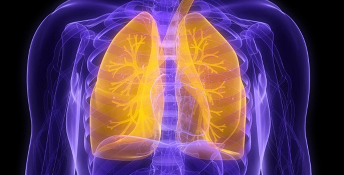
Pulmonary macrophages are a heterogeneous population of immune cells that patrol the lungs, ingesting particulates and microbes, and activating inflammation when necessary. During homeostasis, two main subsets of macrophages coexist: resident alveolar macrophages (AMs) and bone marrow (BM)-derived macrophages. Identifying and characterizing these subsets during lung inflammation is challenging due to altered expression of cell surface markers.
Timothy Blackwell, MD, and colleagues have now developed a technique to separately label AMs (via intratracheal injection) and BM-derived macrophages (via tibial injection) with lipophilic fluorescent dyes that insert into cell membranes. They used the approach to track the macrophage subsets and characterize gene expression profiles at different time points after intratracheal injection of the bacterial toxin LPS.
The study, reported in The Journal of Immunology, shows that the new method accurately distinguishes AMs and BM-derived macrophages during LPS-induced inflammation. It should be useful for studying macrophage subsets during lung inflammation, infection and injury, the authors note.
Co-authors of the report included Wei Han, MD, Harikrishna Tanjore, PhD, Yang Liu, PhD, Raphael Hunt, MS, Sergey Gutor, PhD, and Ana Serezani, PhD. The research was supported by the National Institutes of Health (grant HL151016) and the Department of Veterans Affairs.