A study of a unique spatial map of gene expression in 1.6 million cells from the lungs of 35 people with pulmonary fibrosis (PF) revealed a discovery that could eventually change how early PF can be detected. Some lung tissue in these patients shows signs of the disease before significant structural remodeling of the tissue occurs.
The study, co-authored by Vanderbilt University Medical Center’s Jonathan Kropski, MD, the Rudy W. Jacobson Professor of Pulmonary Medicine and associate professor of Medicine and of Cell & Developmental Biology, and a team of VUMC researchers, was published Feb. 3 in Nature Genetics.
The research, conducted along with a team from TGen, part of City of Hope, and St. Vincent’s Medical Research Institute in Melbourne, Australia, could point to future therapeutic strategies that treat PF patients based on their individual stage of cellular and molecular remodeling.
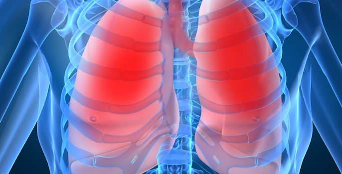
The study, which used image-based spatial transcriptomics to characterize the localization of PF-emergent cell types, establish the cellular and molecular basis of classical PF histopathologic features, and identify a diversity of distinct molecularly defined spatial niches in control and PF lungs, could have far-reaching benefits for patients diagnosed with PF, a chronic lung disease characterized by progressive thickening and scarring of lung tissue.
This scarring reduces lung function over time, causing symptoms such as shortness of breath, persistent cough and fatigue. Current treatments for the most common and severe form of PF only slow declining lung function. Most patients die or require lung transplantation within three to five years after their diagnosis.
“What we are seeing from this work is the cellular processes that go wrong at the beginning of this disease are not necessarily the things that current treatments would be expected to have any effect on,” Kropski said. “As we get a more nuanced understanding of what is driving these changes, we hope these will lead to clues about what pathways we need to nudge the alveoli back to health.”
Image-based spatial transcriptomics, the technique used by the scientists, is an important tool for studying PF. To better view this complex cellular process, the researchers profiled 343 genes in lung tissue samples from 26 people who underwent lung transplant for PF and from nine people without PF.
“We were intentional in designing this study to make sure we had the ability to look at not just healthy and end-stage disease samples, but also those across a spectrum of pathologic remodeling,” said Nicholas Banovich, PhD, from TGen, another of the study’s senior authors.
Their analysis helped them map out where cells with signs of PF occur and identify the cellular and molecular underpinnings of some features of PF. They also characterized 12 distinct, molecularly defined spatial niches in healthy and PF-affected lungs.
“We tried to dig down into this question: How does the disease evolve?” Kropski said.
“We developed an innovative machine learning-based method to segment the tissue samples into individual alveoli and then order those alveoli by the degree to which they expressed disease-associated molecular profiles to recreate the spectrum from normal to a bit dysregulated to more advanced and end-stage disease. That gave us the opportunity to determine which cells show changes the earliest — to chart the ‘molecular order of operations’ on how we think disease unfolds, at least on a cellular basis,” said Jennifer Sucre, MD, associate professor of Pediatrics and one of the study’s co-investigators.
VUMC and TGen researchers will continue to expand their samples and look more closely at the mechanisms behind early PF in three research collaborations.
“We are excited that these findings give us hope that we may be able to halt or reverse disease by targeting early molecular changes before patients suffer substantial loss of lung function and develop debilitating symptoms from pulmonary fibrosis,” Kropski said.
Other members of the VUMC research team are: Nicholas Negretti, PhD, Joseph Hirsh, Samuel Hirsh, David Nichols, Carla Calvi, Chase Taylor, Vasiliy Polosukhin, MD, PhD, Ana Serezani, PhD, Scott McCall, MD, PhD, Jason Gokey, PhD, Lorraine Ware, MD, Matthew Bacchetta, MD, and Ciara Shaver, MD, PhD.
The National Institutes of Health, Francis Family Foundation, Vanderbilt Faculty Research Scholars, the Department of Veterans Affairs, and the National Health and Medical Research Council funded this study (U01HL175444 and R01HL145372).