For the first time, a fluorescent-guided nerve imaging agent shows promise for use in humans, according to a paper published in Nature Communications. The study sought to evaluate the safety of bevonescein, a synthetic peptide-dye conjugate thought to be applicable for intraoperative nerve-specific fluorescence imaging.
Eben Rosenthal, MD, chair of the Department of Otolaryngology-Head and Neck Surgery, served as the paper’s senior and corresponding author, and Sarah Rohde, MD, MMHC, division chief of Head and Neck Surgery, is the project’s lead investigator for Phase 3 clinical trials. Their findings, published with and associates from other centers around the country, identified a safe and effective fluorescent imaging agent that can assist surgeons in visualizing — and therefore protecting — nerves during surgery.
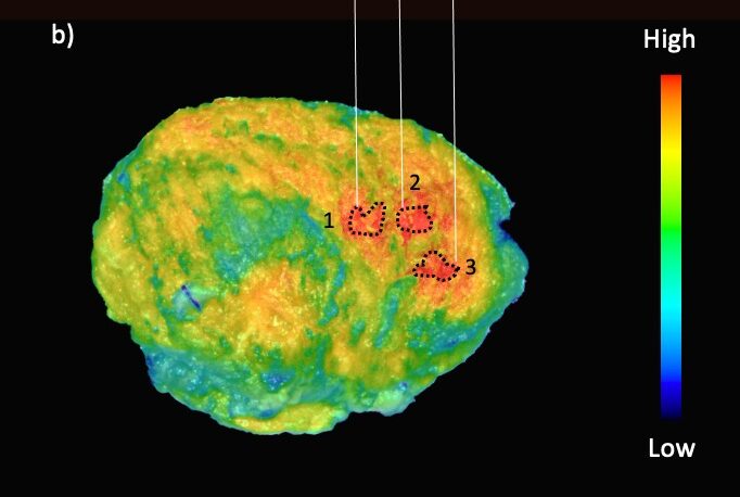
Through intravenous infusion of a fluorescent imaging agent such as bevonescein, surgeons can use excitation light to illuminate a patient’s tissue during surgery to guide their incisions, thus making it easier to locate and avoid injuring nerves. While previous research into nerve-specific fluorescence imaging had only yet been successful in animals, the finding that bevonescein is the first such agent safe for use in humans is a significant breakthrough in intraoperative safety for head and neck surgery.
“Nerve injury is a major problem related to surgical intervention. The idea is that if you can see nerves better, you would avoid injuring them,” said Rohde, who helped Vanderbilt recruit the greatest number of patients in the study. “Different technologies have all come together over the past 10 years to allow the development of intraoperative molecular imaging, or fluorescent-guided surgery. [We’ve identified] the first in-human nerve imaging agent that shows promise.”
This latest research into nerve imaging, conducted with lead author Yu-Jin Lee, MD, of Stanford University and Alume Biosciences Inc., builds on years of Rosenthal’s success in using fluorescence imaging to target tumors in oncological head and neck surgery at VUMC. Fluorescence imaging for cancer is an integral part of guiding surgical operations and assessing results.
“Cancer is a surgical disease,” said Rosenthal, who holds the Barry and Amy Baker Chair in Laryngeal, Head and Neck Research. “Most cancers get surgery. Primarily early-stage disease … is a cancer that can be taken out and cured with surgery. “[With fluorescence imaging,] you can see the cancer light up, which makes it easier to treat.”
Without fluorescence imaging, though, it can be somewhat of a guessing game to determine exactly where the cancer is closest to the incision.
“We can’t tell where the cancer’s closest because we’re just kind of feeling, but if it has the highest fluorescent intensity, that’s where it’s going to be. It shows up brighter if it’s closer to the surface,” said Rosenthal.
In certain environments, including both cancer and nerve imaging, the use of a fluorescent agent is critical, but in other instances, it may not be helpful. In other words, it doesn’t have universal application — but it doesn’t need to.
“Robotic and laparoscopic [environments] work better, because there’s no ambient light because it’s all within a cavity. It’s a low-light environment,” said Rosenthal. “There are some circumstances where it’s really valuable, and there are others where it’s not. And that’s one of the key things about understanding this process.”
Rosenthal said the ability to apply this technology to both tumors and nerves highlights multidisciplinary success. And with the research on nerve-specific fluorescence imaging now in Phase 3 trials, he’s optimistic about the prospect of getting this method for nerve imaging in front of the Food and Drug Administration for approval in the near future.
“Vanderbilt has been leading in fluorescence imaging … and nerve imaging is a good partnership with our cancer imaging program,” said Rosenthal. “It’s this combination of different technologies coming together at the same time.”
The research was supported in part by the National Institutes of Health (grants R01CA266233 and R37CA245157) and by Alume Biosciences, with which the Vanderbilt authors have no financial conflict of interest.