

Vanderbilt Health and Vanderbilt University researchers in a highly productive collaboration are engineering nanobodies, tiny fragments of unique antibodies produced by alpacas, to probe the mysteries of Alzheimer’s disease.
In 2024 they reported that nanobodies labeled with a fluorescent dye cross the blood-brain barrier and “light up” toxic aggregates of amyloid proteins in the synapse, the tiny gap between nerve cells that enables neurons to communicate with each other.
In recent work, the researchers discovered a nanobody that neutralized these small synaptic aggregates in the hippocampus, a part of the brain important in memory and learning.
An Australian biotechnology company has now licensed their nanobody invention for further development and exploration of its “theragnostic” potential, both in diagnostic imaging and as targeted therapy to prevent or slow the progression of the disease.
Nanobodies are “a powerful tool to investigate the potential mechanism of synaptic dysfunction,” which could be the initial step leading to neuronal death and tissue atrophy in Alzheimer’s disease, said Wellington Pham, PhD, a leading expert in molecular probe design at Vanderbilt.
Pham, professor of Radiology and Radiological Sciences and Biomedical Engineering, co-led the study with Brian Wadzinski, PhD, and Ben Spiller, PhD, associate professors of Pharmacology. The study was undertaken in 2020, at the height of the COVID-19 pandemic, and successfully completed with support from multiple research institutes at Vanderbilt.
According to long-held theory, Alzheimer’s disease arises from the accumulation of amyloid plaques and neurofibrillary tangles that kill nerve cells. Recent evidence suggests, however, that soluble amyloid-beta oligomers (SAβOs), toxic aggregates of protein fragments, accumulate in the synapse prior to plaque formation and disrupt cognitive function.
The project began in an “outdoor laboratory” of alpacas in rural Humphreys County, Tennessee, that supports nanobody research at Vanderbilt and around the country. One of the animals was immunized with an antigen, a protein marker of the SAβO molecule, to stimulate an immune response against it.
Two months later, the researchers collected blood samples from the animal and from its unique, “heavy-chain” antibodies, used molecular engineering techniques to isolate and amplify the amino acid sequence of an antibody fragment that specifically binds SAβO.
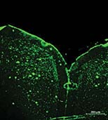
The resulting nanobody, when labeled with a fluorescent dye, illuminated the precise locations of SAβO in brain tissue from a mouse model of Alzheimer’s disease. This suggests that, when injected intravenously, the nanobody could enable “precision in vivo molecular fingerprinting of SAβOs for early detection,” the researchers concluded.
The researchers acknowledged the support and assistance provided by the Vanderbilt University Institute of Imaging Science, Vanderbilt Brain Institute, Vanderbilt-Ingram Cancer Center, and Vanderbilt Institute for Clinical and Translational Research.
The project was partially supported by a grant from the National Institute on Aging (R01AG061138).