



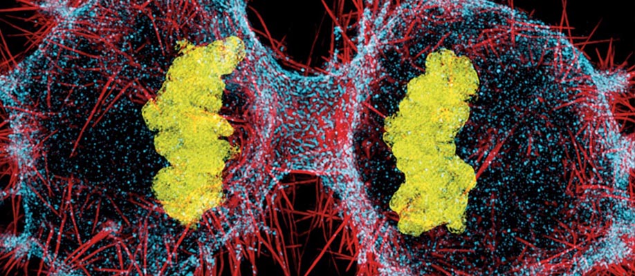

Dylan Burnette, PhD, assistant professor of Cell and Developmental Biology at Vanderbilt, uses a structured illumination microscope to capture cancer cells in motion. This advanced microscope shines different patterns of light on a sample to reveal images that are blurred by older microscope designs. The microscope he and other researchers use is housed at the Vanderbilt Cell Imaging Shared Resource, which is a Nikon Center of Excellence, one of only 21 such centers worldwide.
Featured Story
January 3, 2019