Richard Caprioli Archive — Page 1 of 1

Vanderbilt CTTC logo with the text “Center for Technology Transfer & Commercialization.”
September 15, 2023

Four named Master Innovators at Vanderbilt

Richard Caprioli, C. David Weaver, Susan Eagle, and Franz Baudenbacher as Master Innovators at Vanderbilt.

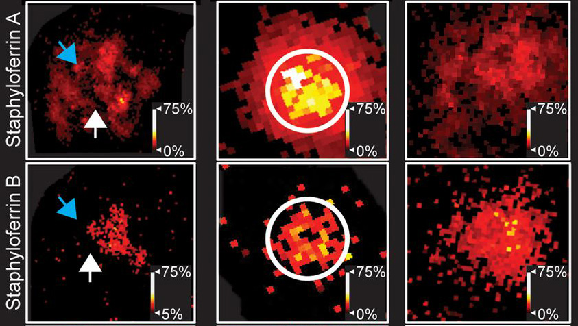
Six-panel fluorescence microscopy figure comparing Staphyloferin A (top row) and Staphyloferin B (bottom row), with red-to-yellow intensity maps, arrows, circled regions, and percentage color scales.
October 31, 2019

Imaging host-pathogen battle for metal

An unprecedented view of bacterial products within infected tissues opens new opportunities to explore infection biology and devise novel therapeutic strategies.

Four researchers pose in front of a colorful imaging mass spectrometry image of a mouse kidney infected with Staphylococcus aureus; the image shows distinct protein species in different colors and spatial distributions.
March 15, 2018

New imaging approach offers unprecedented views of staph infection

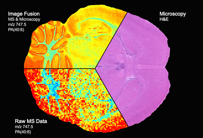
A new integrated imaging approach makes it possible to probe the molecules involved in invasive infections and can be broadly applied to any health or disease state.

Brain section divided into three panels: a pink microscopy image labeled “Microscopy H&E,” a colorful fused mass-spectrometry and microscopy image labeled “Image Fusion MS & Microscopy m/z 747.5 PA(40:6),” and pixelated raw mass-spectrometry data labeled “Raw MS Data m/z 747.5 PA(40:6).”
March 5, 2015

Vanderbilt team first to blend high-end imaging techniques

Vanderbilt University researchers have achieved the first “image fusion” of mass spectrometry and microscopy — a technical tour de force that could, among other things, dramatically improve the diagnosis and treatment of cancer.

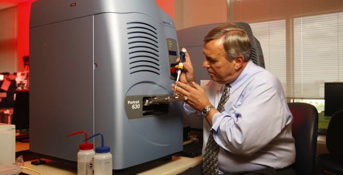
Richard Caprioli and mass spectrometer
March 3, 2014

Vanderbilt awarded $16.5 million agreement to determine how toxic agents affect human cells

Vanderbilt University has been awarded a Cooperative Agreement with the Defense Advanced Research Projects Agency and the Army Research Office that is worth up to $16.5 million over five years.

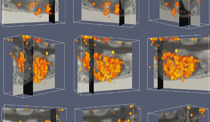
Grid of grayscale 3D medical imaging renderings with orange and yellow highlighted regions shown in transparent box views.
August 9, 2012

Team creates new view of body’s infection response

A new 3-D view of the body’s response to infection — and the ability to identify proteins involved in the response — could point to novel biomarkers and therapeutic agents for infectious diseases.