
Vanderbilt University Institute of Imaging Science (VUIIS) Archive — Page 1 of 6

-
February 7, 2024
Vanderbilt nanodrug may be a paradigm shift for cancer
A multidisciplinary research team at Vanderbilt University and Vanderbilt University Medical Center has discovered a new way to kill a tumor by disrupting its acidic “microenvironment” without harming normal tissue. -
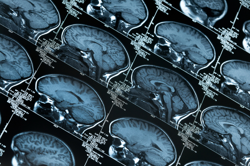
January 30, 2024
3D brain mapping opens a window to the aging brain
By mapping brain activity in three dimensions, researchers at Vanderbilt University Medical Center have achieved a more detailed picture of how the brain changes with age. -
December 20, 2023
Nonaddictive pain relief system nears clinical trials
Researchers in the Vanderbilt are nearing completion of an ingenious undertaking that may be a highpoint of their careers — a non-addictive alternative for relieving chronic pain. -
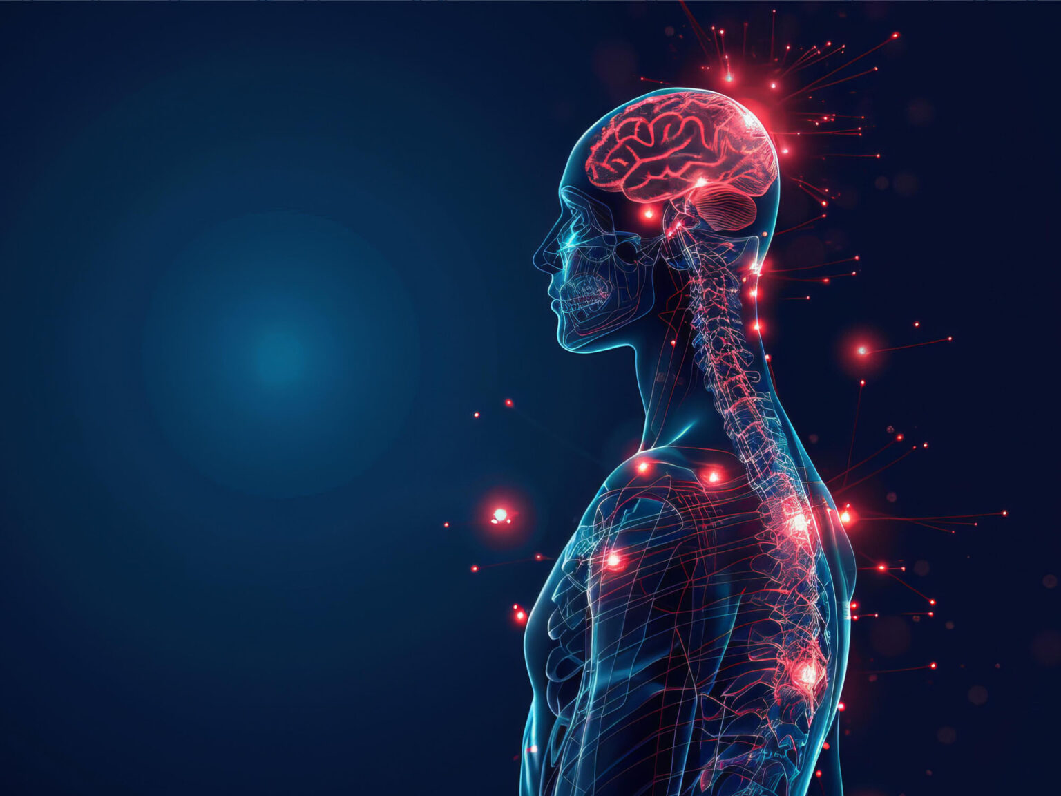
October 16, 2023
VUMC scientists record powerful signal in the brain’s white matter
Vanderbilt researchers report that when people who are having their brains scanned by fMRI perform a task, like wiggling their fingers, certain signals increase in white matter throughout the brain, which has long been thought to play a lesser role the more the brain's more energetic gray matter. -
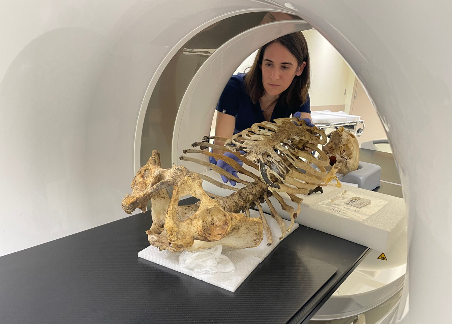
June 8, 2023
An image of cancer treatment response
An MRI method developed by Vanderbilt researchers uses imaging data to derive cell size and could be added to clinical practice for better monitoring of breast cancer treatment response. -
May 8, 2023
Improved imaging for kidney disease
Vanderbilt researchers identified optimal MRI parameters for estimating the severity of polycystic kidney disease, a common inherited disorder that can lead to end-stage renal failure. -
November 3, 2022
New VUSM master’s program offers broad training in biomedical imaging
A new Master of Imaging Science program at Vanderbilt will immerse students in the full spectrum of biomedical imaging and provide hands-on clinical and research experience.