Functional magnetic resonance imaging (fMRI) has become the leading technique for mapping neuronal activities in the brain by detecting changes in blood-oxygen-level-dependent (BOLD) signals.
Power spectra analysis — a plot of signal power against frequency — has been used to determine differences in BOLD signal changes across different areas of gray matter, where most of the neuronal cell bodies driving brain functions are located.
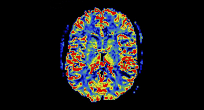
Using power spectra analysis, Muwei Li, PhD, and colleagues in the Vanderbilt University Institute of Imaging Science discovered intriguing patterns of BOLD signals in white matter, which consists of the nerve fibers and their protective myelin coating that are essential for relaying signals between different brain regions.
In particular, they found distinct time courses in BOLD signals during a resting state that may reflect different anatomical structures and functional connectivities within the tissue.
These findings, published in the Proceedings of the National Academy of Sciences, provide novel insights into the fundamentally unique structural-vascular-functional association in white matter and its role in overall brain function.
The research was funded in part by National Institutes of Health grants NS093669 and NS113832. Data provided by the Human Connectome Project, Washington University-University of Minnesota Consortium.