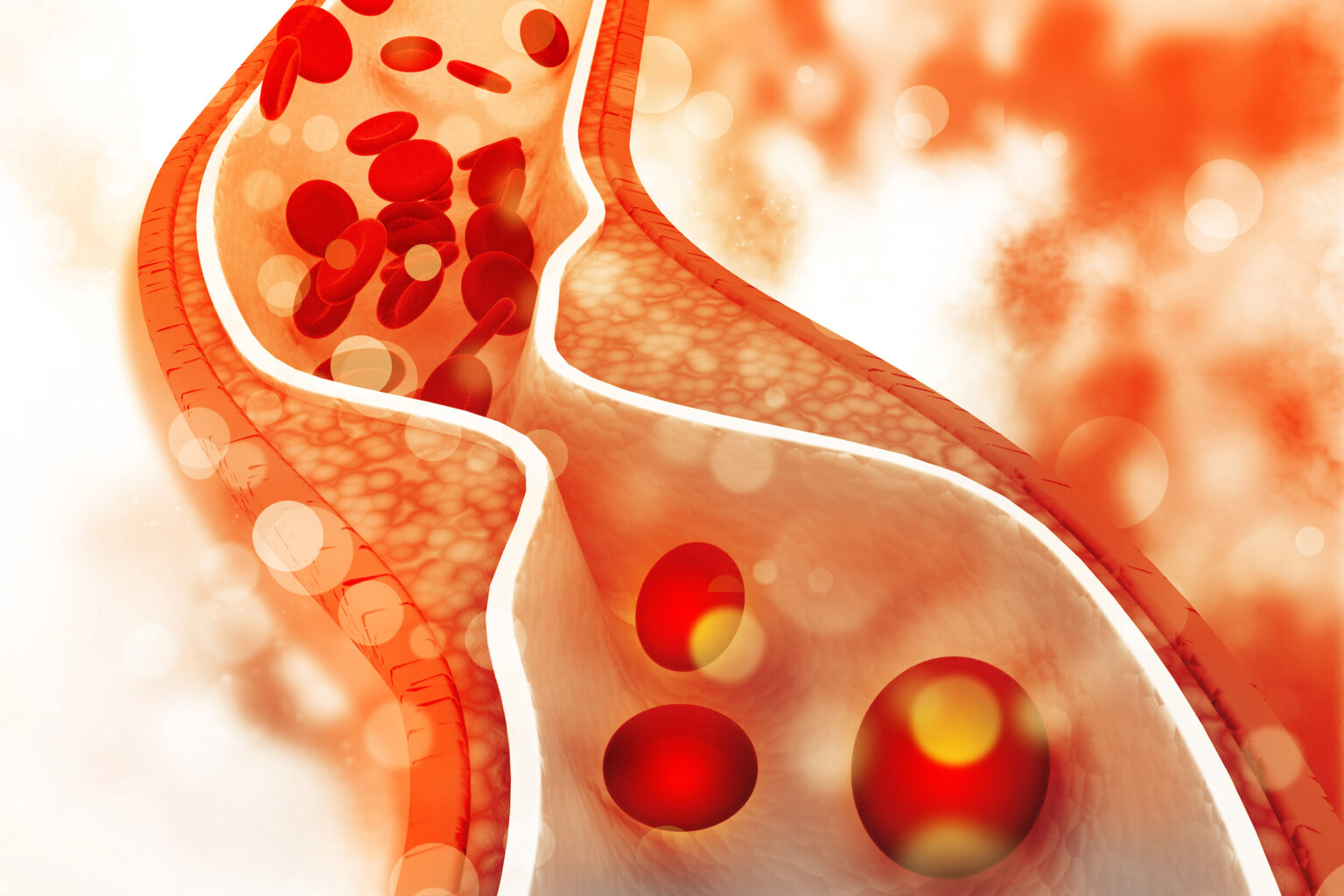
Atherosclerosis is the slow and progressive narrowing of arteries due to plaque formation. The atherosclerotic plaque forms by local proliferation of leukocytes and vascular smooth muscle cells (VSMCs) in the vessel wall along with associated changes in cellular metabolism.
Until now, it was technically impossible to study the changes in cell metabolism and proliferation of cells directly in atherosclerotic plaques at the same time.
Reporting in the journal JCI Insight, Jonathan D. Brown, MD, and colleagues for the first time have applied a quantitative imaging method called multi-isotope imaging mass spectrometry (MIMS) to directly measure changes in cell division and simultaneous glucose utilization in the atherosclerotic plaques at suborganelle resolution.
Their study, conducted with colleagues at Brigham and Women’s Hospital in Boston, demonstrates significant heterogeneity in VSMC glucose metabolism that varies both with proliferative status and proximity to growing plaques, and establishes MIMS as a powerful, complementary approach to studying the cell biological processes of atherosclerosis in vivo.
This research was supported by grants from the National Institutes of Health (HL116263, HL127173, DK59637) and by a VICC Ambassadors Award.