Immune system T cells that should be able to kill cancer cells become dysfunctional or “exhausted” within hours of encountering a tumor, according to a study reported Aug. 3 in Nature Immunology.
The surprising findings have implications for cancer immunotherapies that aim to harness the tumor-killing power of T cells, and they challenge existing ideas about how T cells become exhausted, said Mary Philip, MD, PhD, assistant professor of Medicine in the Division of Hematology and Oncology at Vanderbilt University Medical Center.
“The idea has been that T cells that are exposed to an antigen (such as a tumor or pathogen) for a long time are working and working, and then at some point they sort of peter out. That’s where the term exhaustion comes from,” said Philip, senior author of the study.
“I don’t think anyone expected that within six to 12 hours, T cells would look dysfunctional or exhausted; that’s a very fast time window.”
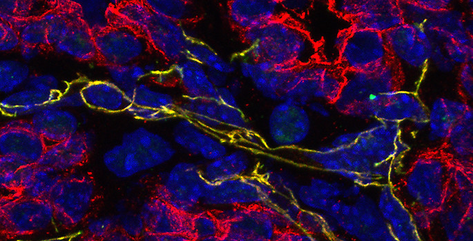
To explore how T cells become exhausted and identify targets that could prevent or reverse it, Philip and her colleagues used their previously established genetic mouse model, in which the mice develop liver tumors as they age in a manner analogous to human patients. The researchers can track immune responses as tumors develop, and they can introduce trackable T cells and study how the cells respond to established tumors.
“In patients who are diagnosed with cancer, we can’t go back in time to find out how the immune system responded,” Philip said. “The mouse model allows us to do this, to say, ‘What is happening when T cells first see tumors; when and how do the T cells get exhausted?”
In parallel studies, the researchers are able to compare how T cells respond to tumors versus infection. In the context of acute infection, T cells become functional, whereas in the context of tumors, they become dysfunctional, Philip explains.
“This comparison allows us to compare what a “good” T cell looks like versus a dysfunctional T cell,” she said.
In a previous set of studies published in 2016 and 2017, the researchers were surprised to find that after only five days, tumor-activated T cells were dysfunctional and had thousands of differences in genes that are turned on or off, compared to infection-activated T cells.
In the current studies, led by Michael Rudloff, PhD, a student in the Medical Scientist Training Program, the researchers looked at earlier times. They hoped they would find fewer differences between infection-activated and tumor-activated T cells, potentially pointing the way to key genes or signaling pathways that could be targeted to make the dysfunctional T cells “good” again. Instead, they found all the hallmarks of T cell exhaustion within six to 12 hours, including dramatic changes in chromatin accessibility and gene expression.
“We still have thousands of different genes to look at,” Philip said.
Despite not finding a few key targets, the researchers found certain trends, such as increased expression of inflammation-associated genes and transcription factors during infection, which are not turned on in the tumor model. They are now exploring infection and other ways of activating innate immune pathways to boost T cell function.
Other key findings of the current study:
- T cells activated and divided after tumor exposure, but differences in function were evident before any cell division, which was unexpected.
- T cells in a metastatic melanoma model also became dysfunctional within hours, demonstrating that the changes do not depend on tumor type.
- T cells that were isolated from the tumor after five days or more and transferred to a tumor-free mouse were not able to regain function, suggesting that the changes are “imprinted.”
- Fully activated and functional T cells introduced into the tumor model became dysfunctional, showing that negative tumor signals can override functional programs.
The researchers have been able to use data from these and other studies to identify biomarkers that allow them to predict whether T cells will respond to a tumor or not, which could help personalize immunotherapies.
“We know that checkpoint inhibitors and other immunotherapy interventions don’t work in a lot of patients, and it’s important to be able to predict response and avoid therapies that won’t benefit patients,” Philip said.
Rudloff is the first author and Philip is the corresponding and senior author of the Nature Immunology paper; co-authors include Paul Zumbo, Friederike Dündar, PhD, and Doron Betel, PhD, from Weill Cornell Medicine, and Natalie Favret, Jessica Roetman, PhD, Carlos Detrés Román, Megan Erwin, Kristen Murray and Sriya Jonnakuti in the Division of Hematology and Oncology and Department of Pathology, Microbiology and Immunology at VUMC.
The research was supported by a V Foundation Scholar Award, grants from the National Institutes of Health (CA263614, CA098131, DK058404, GM007347, GM008554, CA009592, AR059039), and the Serodino Family Adventure Allee Fund.