


cell and developmental biology Archive — Page 2 of 9




-
September 8, 2016
Going after the ‘heart attack gremlin’
A protein called Gremlin 2 controls the extent of inflammation after heart attack and may be a good therapeutic target. -
July 20, 2016
Breast cancer: finding the smoking gun
A new method developed at Vanderbilt may help “inventory” all tumor-promoting genes. -
June 23, 2016
It takes two to tango: beta cell development
Defining the genes required for the function of insulin-producing beta cells is crucial for ongoing efforts to develop a cell-based therapy for diabetes. -
June 2, 2016
‘Young Scientist’ showcases high schoolers’ research at Vanderbilt
High school students performing advanced research at Vanderbilt have the opportunity to share their findings with the scientific community through a journal of their own. -
April 28, 2016
An Argonaute’s voyage to cancer
A genetic mutation that promotes cancer development blocks the normal sorting of a protein called “Argonaute 2.” -
April 28, 2016
Zanic’s research recognized with 2016 Searle Scholarship
An expert in the dynamic behavior of the microtubule cytoskeleton at Vanderbilt University is among 15 scientists in the chemical and biological sciences nationwide who have been named 2016 Searle Scholars. -
April 21, 2016
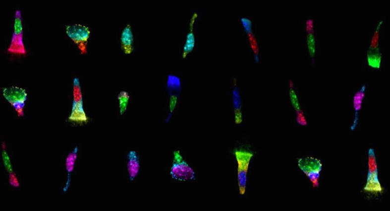
Nobel laureate Betzig set for Discovery Lecture
Eric Betzig, Ph.D., who shared the 2014 Nobel Prize in Chemistry for the development of super-resolved fluorescence microscopy, will deliver the next Flexner Discovery Lecture on April 28.