A team of investigators led by Fabien Maldonado, MD, associate professor of Medicine at Vanderbilt, and Tobias Peikert, MD, assistant professor of Medicine at Mayo Clinic, Rochester, Minnesota, has identified a new technology to address false positives in CT-based lung cancer screening. The study was published in the latest issue of PLOS One.

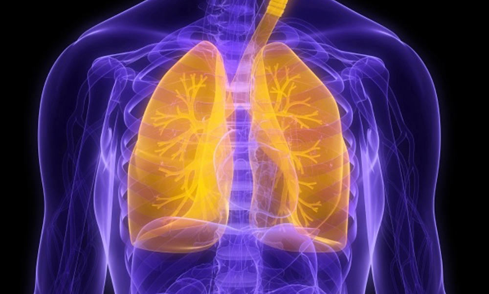
Lung cancer is the leading cause of cancer-related death in the United States, claiming approximately 160,000 patients annually. The disease is lethal because it is often found at late stages when it is not curable.
In 2011, the National Lung Screening Trial (NLST) demonstrated a 20 percent relative reduction in lung cancer mortality with the use of annual low-dose computed tomography (CT) screening among high-risk patients aged 55 to 74. As a result of this study, lung screening programs have been widely adopted and VUMC’s Lung Screening Program recently reached a milestone, enrolling more than 700 patients and performing more than 1,000 CT screening examinations.
While the nation’s screening programs have identified at least one pulmonary nodule in nearly 40 percent of patients, 96 percent of those nodules are eventually found to be benign.
“This high rate of false positives creates a dilemma for physicians and patients because patients may be subjected to unnecessary tests and procedures that may have risks,” said Maldonado, who also serves as associate professor of Thoracic Surgery and Mechanical Engineering. “The need for additional testing also adds to the nation’s health care costs.”
To address the problem of false positives in lung cancer screening, Maldonado, Peikert and colleagues analyzed the CT images of all lung cancers identified during the NLST using a radiomics approach. This involved extracting large amounts of quantitative data from the CT images and using computer programs to identify disease characteristics that may not be visible to the naked eye.
Non-lung cancer controls were selected as a stratified random sample from all participants without a diagnosis of lung cancer during the screen or follow-up periods of the NLST. Cases with more than one nodule were excluded and the investigators did not include other variables such as current smoking status, prior smoking history or age.
The investigators tested a set of 57 variables for nodule volume, density, shape, surface characteristics including curvature, and the texture of the lung tissue adjacent to the nodule. They validated eight characteristics that enabled them to distinguish benign nodules from cancerous tumors. The eight characteristics were not directly linked to nodule size.
The authors note that this automated radiomics approach still needs to be validated by other researchers, but if the newly developed technology is confirmed in further testing, it could be used to more reliably distinguish benign from malignant nodules and could lead to better management of lung screening patients who are found to have nodules.
Other investigators on the team include Srinivasan Rajagopalan, PhD, Ronald Karwoski, Ryan Clay, MD, Richard Robb, PhD, and Brian Bartholmai, MD, Mayo Clinic; Fenghai Duan, PhD, Ziling Qin, MSc, and JoRean Sicks, MS, Brown University of Public Health.
Funding for the research was provided by the Department of Defense Lung Cancer Program (W81XWH-15-1-0110) and VUMC.