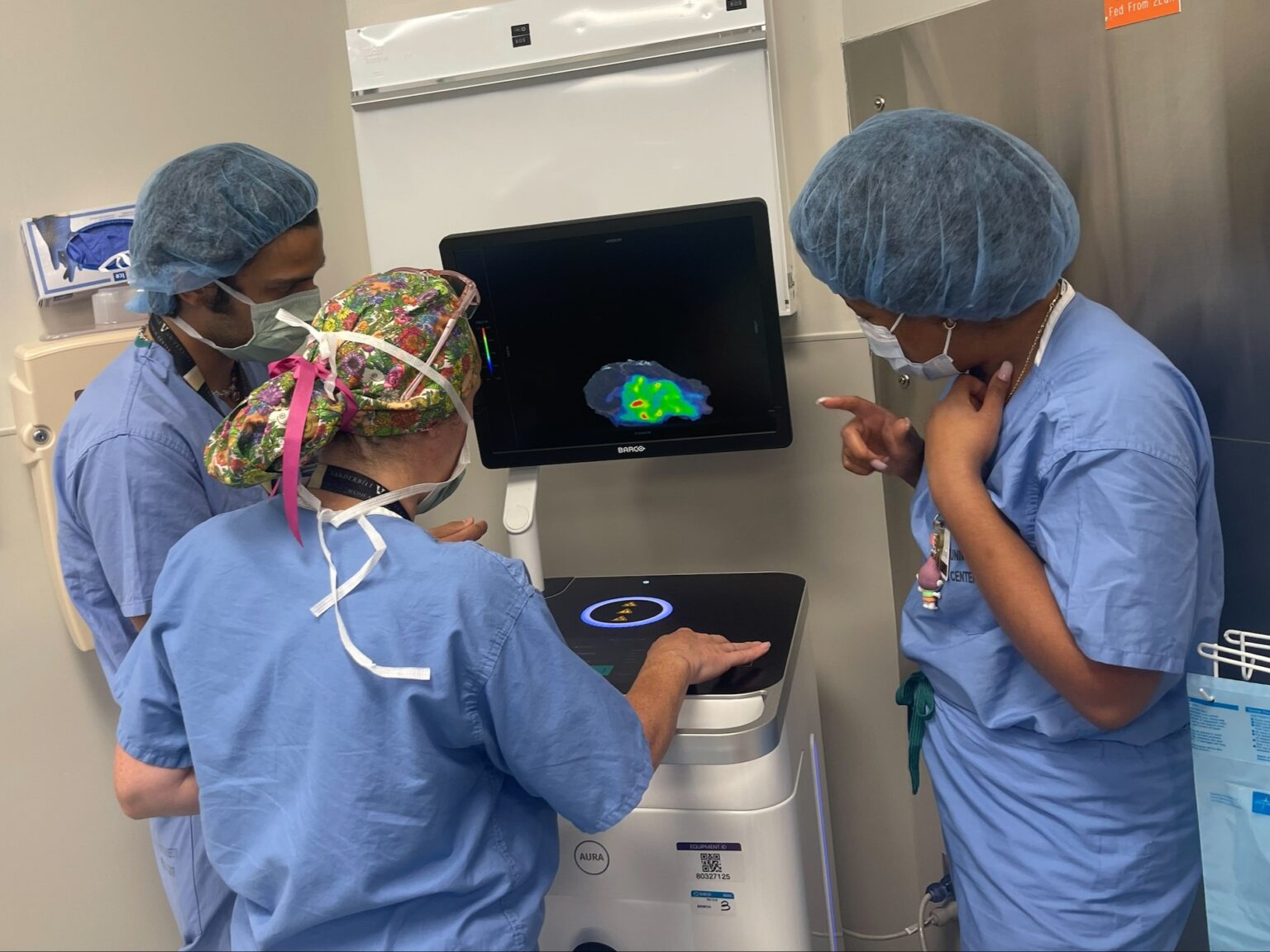
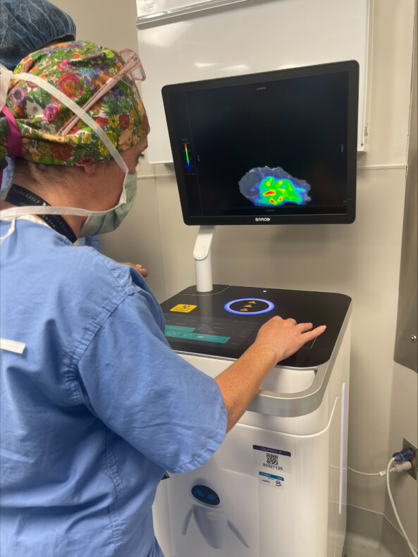
 From left, Andreja Radevic, Denise Garcia, MD, and Kyrionna Golliday review imaging of a breast tumor in the operating room. (photo by Michael Topf)

Following the introduction of a surgical protocol that leveraged intraoperative imaging with a combination PET-CT scanner to assess the success of head and neck cancer resection, surgeons have used the technology for breast cancer.
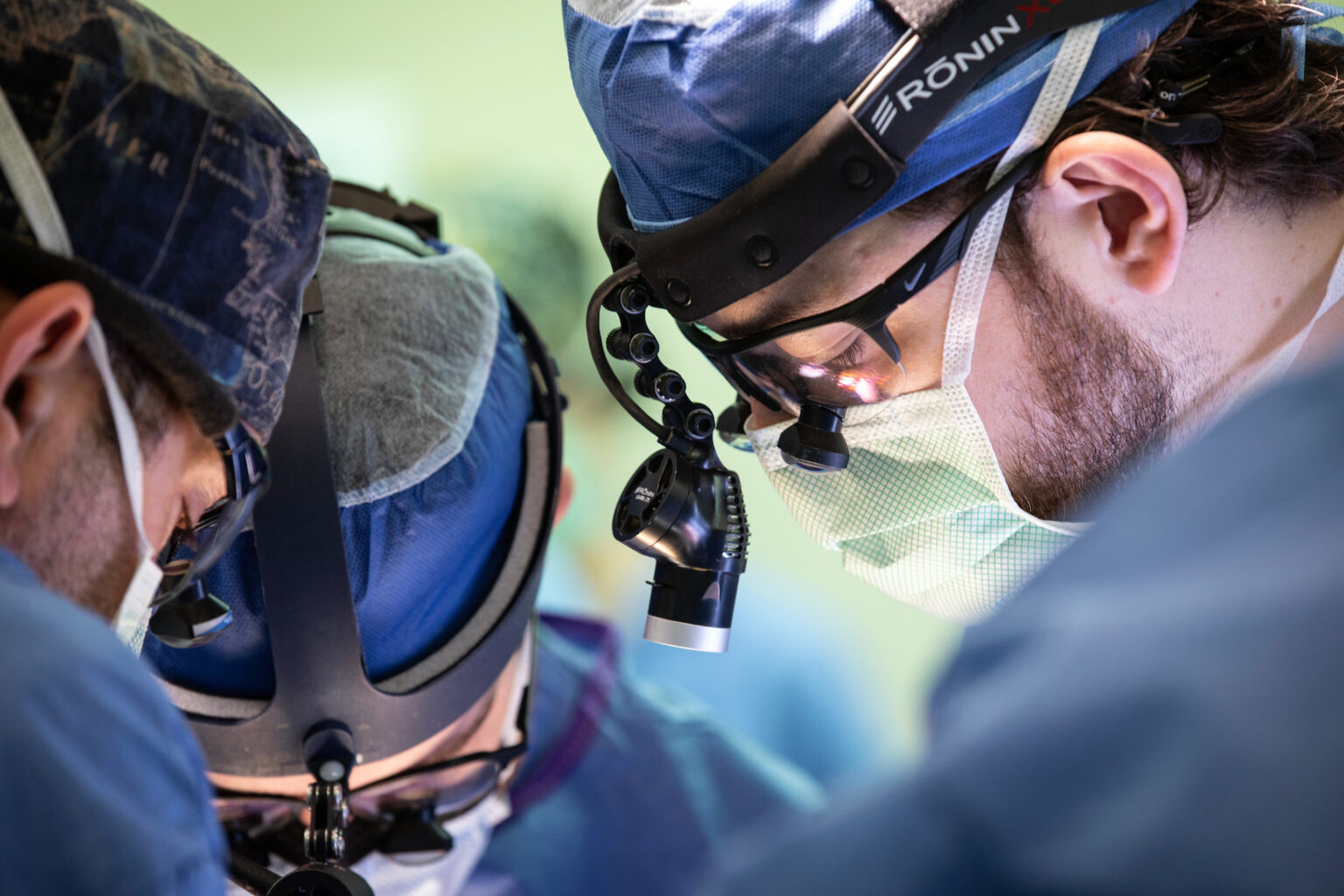
Investigators led by Michael Topf, MD, Associate Professor of Otolaryngology-Head and Neck Surgery, performed the nation’s first surgery using intraoperative PET-CT scanning in September 2025. Now, a surgical team led by Denise Garcia, MD, Assistant Professor of Surgery in the Division of Surgical Oncology and Endocrine Surgery, has applied the technology to successfully resect a breast cancer mass.
“This application of intraoperative PET-CT is proof that countless patients can benefit from the expansion of this novel imaging methodology,” said Garcia. “Our team is proud to apply it to a type of tumor that has not yet been imaged for the purposes of assessing margin status. As our institution expands treatment methodologies to more types of cancer, we can cure more patients and give them peace of mind that their surgery has been completed with precision. This technological advancement underscores the success we’ve had across multiple disciplines in working toward that goal.”
Rapid expansion of intraoperative PET-CT scanning boosts efficiency, offers peace of mind to surgeons and patients alike
With each new application, surgeons are demonstrating that an intraoperative PET-CT imaging protocol can help reduce wait time for results from several days to a matter of minutes and allow surgical teams to know immediately whether they need to continue operating.
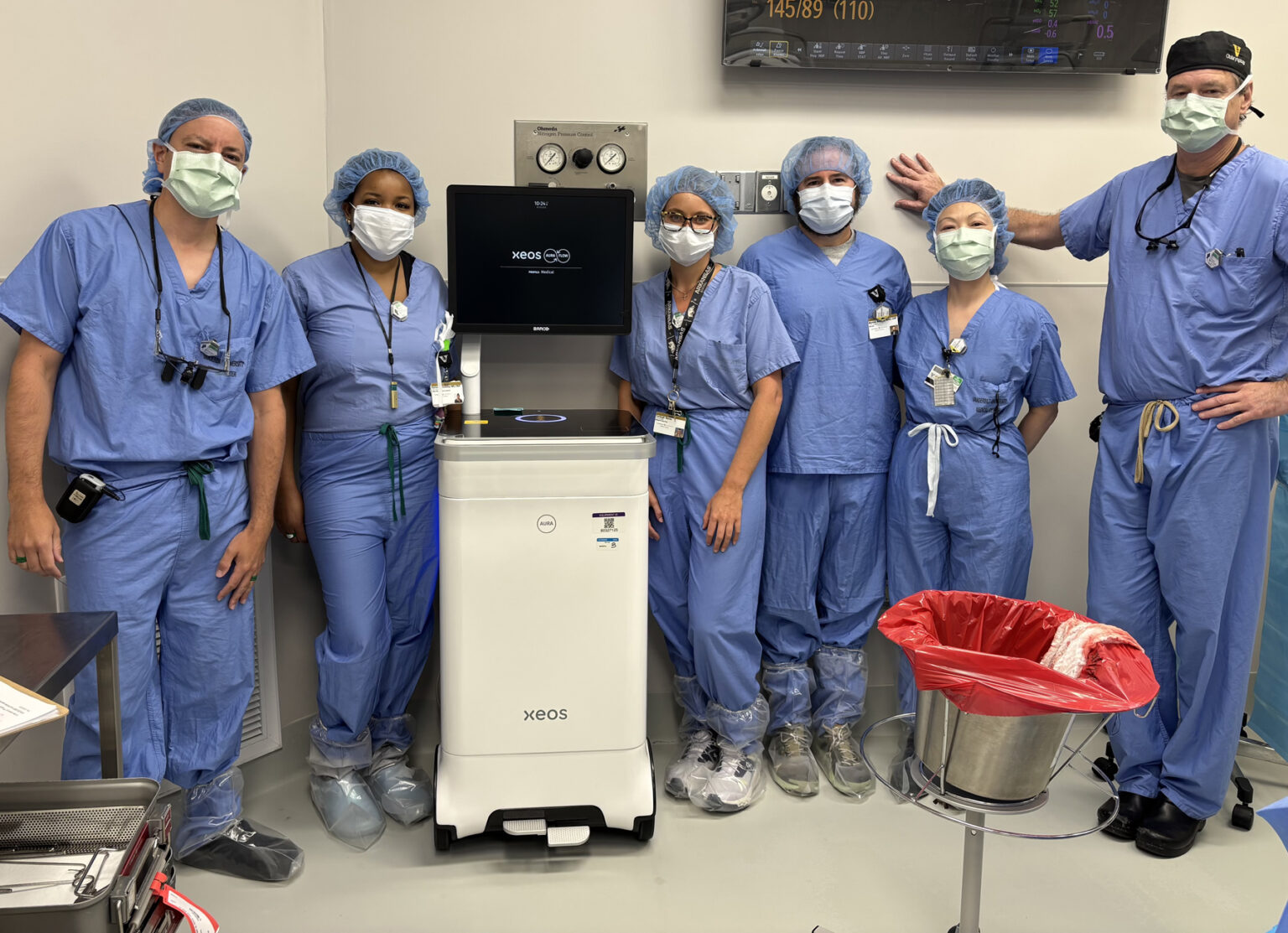
During surgery, the patient receives a dose of a radioactive agent that illuminates the cancer tissue in the scanner. Once the tumor is excised, it is placed in a specialized mobile PET-CT scanner called an Aura 10 device, developed and supplied by Belgium-based surgical technology company Xeos. The scanner negates the need to send the specimen to the pathology lab, providing surgical teams with a real-time view and allowing them to quickly determine if the entire cancerous mass was removed.
If any mass remains, the operation continues. If the cancer has been successfully resected, the surgery concludes, and the patient is sent home with peace of mind, knowing they won’t need to return for a follow-up surgery, and with confidence that their surgeons have a precise, immediate look at the results of the surgery. Because patients receive the radioactive agent on the day of surgery rather than in advance, they also receive a lower dose of radiation.