Jeffrey Spraggins Archive — Page 1 of 1

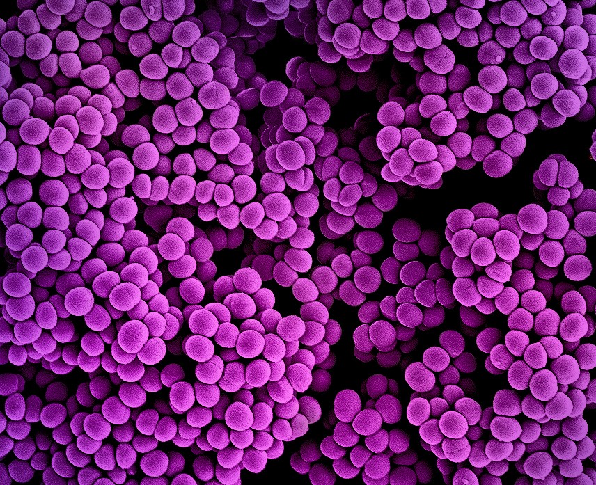
Colorized scanning electron micrograph showing clusters of purple, round MRSA bacteria against a black background.
June 2, 2022

Bacterial battle in 3D

Using imaging mass spectrometry and microscopy, Vanderbilt researchers visualized how staph bacteria modifies lipids in its membrane to evade immune system-mediated killing.

September 30, 2021

Molecular imaging of C. diff infection

C. difficile — the leading cause of hospital-associated intestinal infections — induces a rapid influx of bile acids into the gut, which could provide a novel target for blocking infection.

False-color molecular image of a rounded biological tissue section, with green outer regions, a yellow band, and a pink-purple inner ring surrounding a darker green center. Labels at the bottom identify three molecular signals: m/z 888.5564 3C13 PI(36:4), m/z 888.5711 13C PI(36:3), and m/z 888.6248 SulfoHex-Cer(d18:2/24:0).
October 31, 2019

Vanderbilt investigators lead effort to create map of the human kidney

Short of mandating universal diabetes treatment, regular exercise and low-calorie diets, little can be done to stem the rising tide of kidney failure — unless scientists can figure out why exactly the kidney’s filtration units, the glomeruli, stop working.

Stylized brain scan with a red beam passing through it, surrounded by multiple false-color medical imaging panels.
March 28, 2018

What protein is that?

An improved technology enables high-throughput protein identification in imaging mass spectrometry, aiding proteomics research.