Staphylococcus aureus (“staph”) is an increasingly antibiotic-resistant bacterial pathogen that can cause a variety of life-threatening illnesses.
Researchers at Vanderbilt University School of Medicine have developed integrated molecular imaging techniques that can produce 3D views of the battle between invading pathogens and the body’s immune defenses down to the subcellular level.
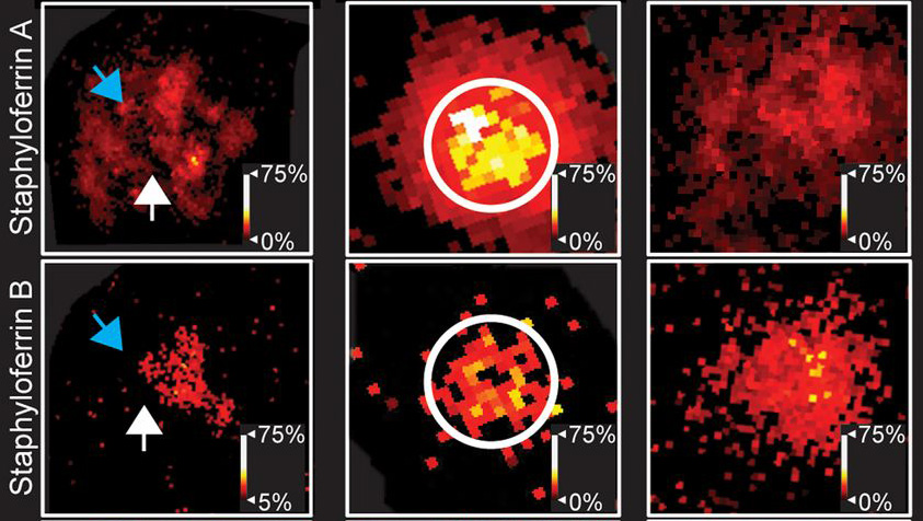
In their latest advance, Jeffrey Spraggins, PhD, Eric Skaar, PhD, and colleagues used multimodal imaging mass spectrometry and microscopy in infected mouse and human tissues to visualize how S. aureus modifies lipids in its membrane that enable it to evade or resist immune-mediated killing.
Distribution of these modified lipids was heterogenous, suggesting that even within the same abscess there are different adaptations or resistant S. aureus subpopulations that can promote chronic infection, the researchers concluded.
These findings, published June 1 in the journal Cell Chemical Biology, are aiding the search for uniformly expressed bacterial factors that could be candidate vaccine targets, as well as inform the development of other novel therapeutics.
The research was supported in part by National Institutes of Health grants GM103391, AI138581, AI145992, AI132560, AI069233, AI073843, and AI150701 and by the Burroughs Wellcome Fund.