
imaging Archive — Page 2 of 3


-
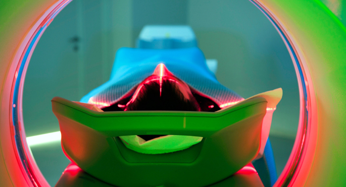
January 31, 2019
Grant supports novel imaging initiative to enhance cancer care
A Vanderbilt initiative to develop predictive imaging technologies that clinicians can use to better match patients with personalized care has received National Cancer Institute (NCI) funding. -
March 15, 2018
New imaging approach offers unprecedented views of staph infection
A new integrated imaging approach makes it possible to probe the molecules involved in invasive infections and can be broadly applied to any health or disease state. -
January 12, 2018
BOLD view of white matter
Vanderbilt investigators have discovered that functional MRI detects neural activity in both gray and white matter in the brain, suggesting new ways to investigate diseases such as Alzheimer’s and multiple sclerosis. -
January 11, 2018
Lighting up iron levels
A new probe enables iron imaging in living animals, providing a unique tool for studying iron’s contributions to health and disease. -
March 30, 2017
Nanobeacon lights up colon tumors
A novel fluorescent nanobeacon can distinguish normal from diseased colon tissue, potentially offering advantages for colorectal cancer screening. -
March 9, 2017
New Radiology website gives patients pre-visit tips
Patients scheduled to undergo an imaging procedure at Vanderbilt University Medical Center (VUMC) can now explore a new Radiology and Radiological Sciences patient website for tips about what to expect during the visit, how to prepare for the procedure and how early to arrive prior to the appointment. -
October 12, 2016
Imaging probe for retinal disease
An imaging probe developed at Vanderbilt detects retinal inflammation early and may allow therapeutic intervention to prevent blindness.