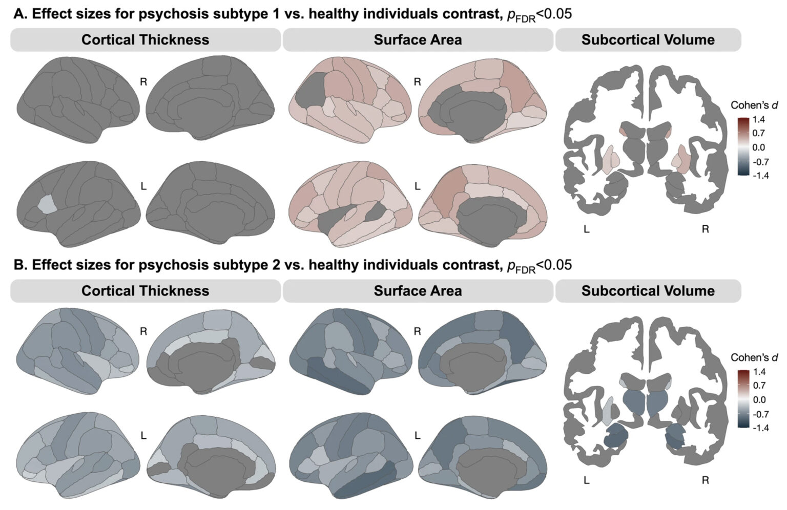
 A. Compared to healthy individuals, psychosis subtype 1 demonstrated significantly larger surface area across the cortex and larger basal ganglia volumes. B. Compared to healthy individuals, psychosis subtype 2 demonstrated widespread lower surface area and cortical thickness, and smaller subcortical volumes. (Hill, L.D., et al. Subtyping psychotic disorders using a data-driven approach reveals divergent cortical and cellular signatures. Mol Psychiatry (2026). https://doi.org/10.1038/s41380-026-03774-2. Creative Commons Attribution 4.0 International License: http://creativecommons.org/licenses/by/4.0/. Rights and Permissions: https://tinyurl.com/2e9nxeah.)

More than 150 years after psychosis was first described in the medical literature, effective treatment options for many patients remain limited.
Now, researchers at Vanderbilt Health have identified two previously unreported subtypes of psychosis based on an analysis of structural brain scans.
Their report, published on July 23 in the journal Molecular Psychiatry, suggests that heterogeneity, or the wide range of symptoms, treatment responses and long-term outcomes observed in patients with various forms of psychosis, may reflect separate illness mechanisms and developmental pathways.
“These data point to the fact that there are different pathophysiological processes that are happening here, and they are at least partially independent,” concluded the paper’s corresponding author, Neil Woodward, PhD, Professor of Psychiatry and Behavioral Sciences.
While further study is needed, “these findings may provide the foundation for more personalized approaches to diagnosing and treating psychiatric disorders,” said Woodward, a clinical neuropsychologist who holds the Charlotte and Donald Test Jr. Chair in Psychiatry.
An estimated 3 million Americans have chronic, debilitating forms of psychosis.
Currently only about a third of patients with psychosis respond well to treatment. “They may never experience another psychotic episode, and if they do, they function relatively well between episodes,” Woodward said.
Another third will follow a relapsing-remitting course, with partial recovery between episodes. The rest have poor long-term outcomes. “They are not very responsive to treatment,” he said. “There are always some residual symptoms, and they have pronounced cognitive impairment.”
It has been known since the 1970s that certain brain structures in patients with psychotic disorders are smaller (have less volume), compared to people who do not have psychosis. Woodward and his colleagues wanted to know if psychosis can be divided into subgroups based on these structural changes.
They analyzed the brain scans of 381 patients who had participated in other neuroimaging studies at Vanderbilt, and who had been diagnosed with either schizophrenia spectrum disorders or bipolar disorder with psychotic features (exhibiting psychotic symptoms only during episodes of mania or depression).
The researchers used an algorithm called similarity network fusion (SNF) to combine measures from structural brain scans, including cortical thickness, cortical surface area, and subcortical brain volumes. This approach enabled them to identify subgroups of patients who shared similar patterns of brain abnormalities.
Replicating prior studies using different methods, SNF sorted the patients into two subgroups. Notably, both subgroups contained individuals with schizophrenia spectrum disorders and bipolar disorder with psychotic features, indicating that structural brain abnormalities do not align with clinical diagnoses.
One subgroup (subgroup 1) showed enlargement of subcortical structures called the striatum in the basal ganglia. Rich in dopamine receptors, selective increase in striatal volume may be attributed to excess dopamine signaling. Most current antipsychotic drugs block dopamine receptors.
Patients in the second subgroup (subgroup 2) demonstrated smaller volumes throughout the brain, including the thalamus and hippocampus, which have been consistently identified as abnormal in psychotic disorders.
Surprisingly, and contrary to the findings of previous brain imaging studies, the two subgroups exhibited very different patterns of changes in the cortex. Subgroup 1 showed moderate enlargement of cortical surface areas, whereas cortical surface area was markedly smaller in subgroup 2.
Individuals in subgroup 2 also exhibited more severe negative symptoms, greater neuropsychological impairment, and lower estimated intellectual functioning compared to those in subgroup 1.
The cerebral cortex governs higher cognitive abilities, including reasoning, problem-solving and speech, as well as voluntary movement and processing sensory information. It begins to develop before birth, grows rapidly through age 2 to 3, and then starts to plateau.
The striking differences in cortical surface area between the two subgroups “suggest there is something that went wrong in very early brain development,” Woodward said. “That’s where these two groups are starting to diverge.”
Using a brain atlas that shows the distribution of different cell types across the cortex, the researchers found that the cortical areas in subgroup 2 with smaller surface area overlap with regions enriched for a specific type of neuron, layer 5 intratelencephalic neurons.
“Based on what we know about these cells, they play a really important role in connectivity between cortical areas that are responsible for higher cognitive function,” Woodward said.
Noting that both subgroups included young people who had experienced their first psychotic episode, Woodward said he hopes to follow the research participants over time.
“If you are subtype 1, do you have a better outcome?” he asked. “Are you more responsive to medication? Does your cognition stay relatively normal?”
Lauren Hill, PhD, Research Instructor in Psychiatry and Behavioral Sciences, is the paper’s first author. Other Vanderbilt co-authors include Baxter Rogers, PhD, Anna Huang, PhD, Victoria Fox, MA, CPA, Brandee Feola, PhD, and Stephan Heckers, MD, MSc.
The research was supported by the National Institutes of Health (grants R01MH102266, K24MH126280, P50MH132642-5980, R01MH070560 and R01MH120080), the Charlotte and Donald Test Fund, and the Vanderbilt Institute for Clinical and Translational Research.